
|
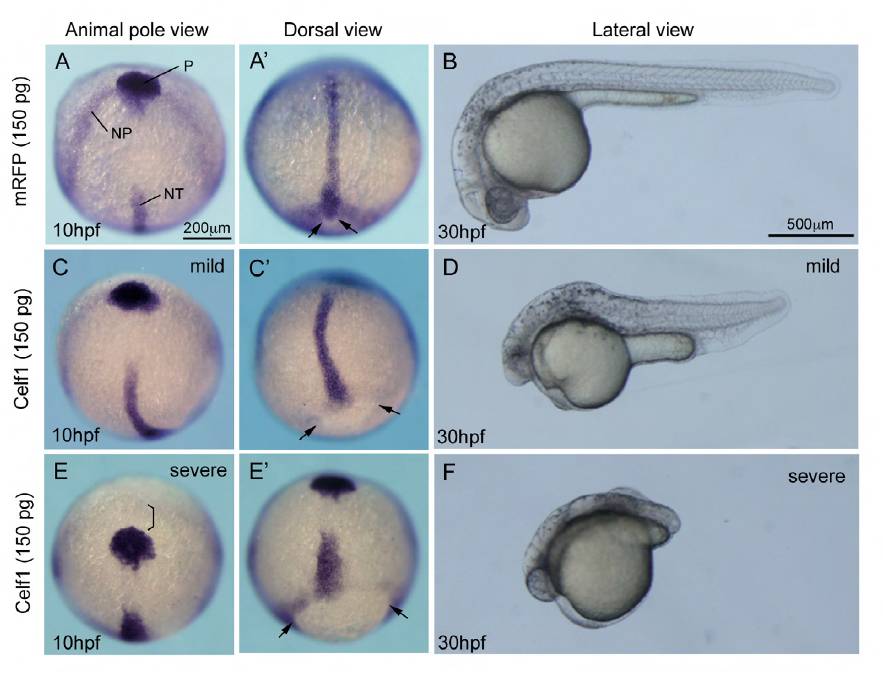
Fig. S4 Higher amounts of celf1 mRNA lead to additional defects in zebrafish. (A,C,E) Expression of hgg1 (P, polster), dlx3 (NP, anterior edge of the neural plate) or ntl (NT, notochord) in embryos injected with mRFP (A) or celf1 (C,E) mRNAs (150 pg). Animal pole view. Scale bar: 200 μm. (A′,C′,E′) Dorsal view of the embryo, anterior to the top. Injection of 150 pg celf1 mRNA resulted in the formation of bended (C) or short (E′) notochord. The polster did not reach the anterior edge of the neural plate (bracket in E). Epiboly was incomplete (C′,E′). Arrows in A′, C′ and E′ mark edge of the yolk plug. (B,D,F) Lateral view of embryos injected with mRFP (B) or celf1 (D, F) mRNAs (150 pg) at 30 hpf. Various phenotypes, such as short tails, segmentation defects, small eyes, small heads and less pigmentation, were observed (D,F).