
|
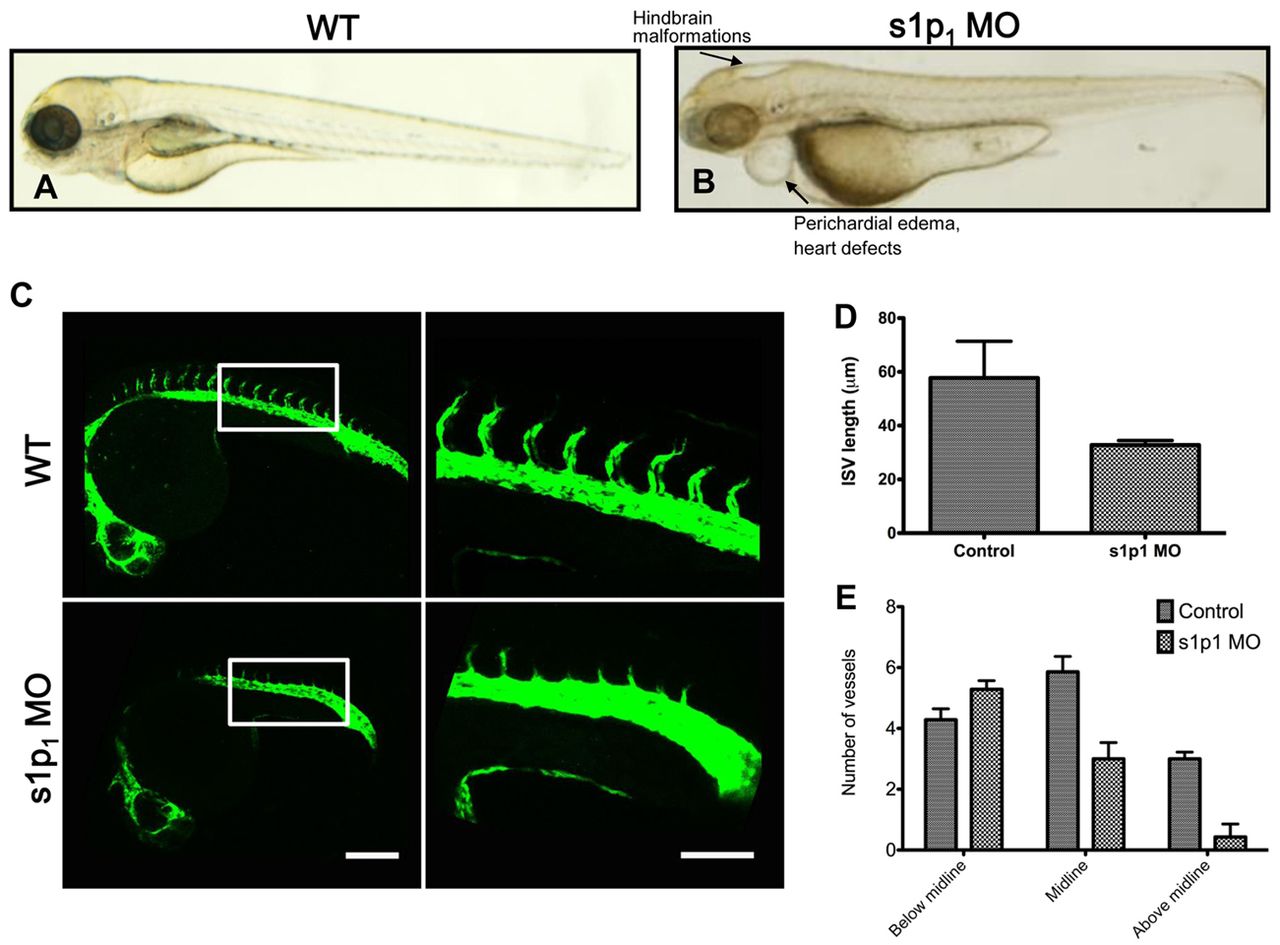
Fig. 5
s1p1 knockdown in zebrafish causes defects in blood vessel formation. Tg(fli-egfp)y1 zebrafish embryos were injected with a control MO and a translation-blocking s1p1 MO and analyzed at 3 dpf. (A,B) Bright-field images of control and s1p1 MO embryos. Arrows in B indicate hindbrain malformations, pericardial edema and heart defects observed in the mutant. (C) Fluorescence microscopy of control and s1p1 MO embryos at 24 hpf. Right-hand panel shows magnifications of the boxed areas on the left. (D) Measurements of ISV length (corresponding to the boxed area in C) in control and s1p1 MO embryos (n=8 WT and 8 MO embryos, P<0.0001, error bars represent s.e.m.). (E) Quantification of ISV location relative to the midline (n=10 WT and 10 MO embryos; 6-8 ISVs from each embryo were analyzed, P<0.05, error bars represent s.e.m.). Scale bars: in C, left panel, 200 μm; in C, right panel, 100 μm.