
|
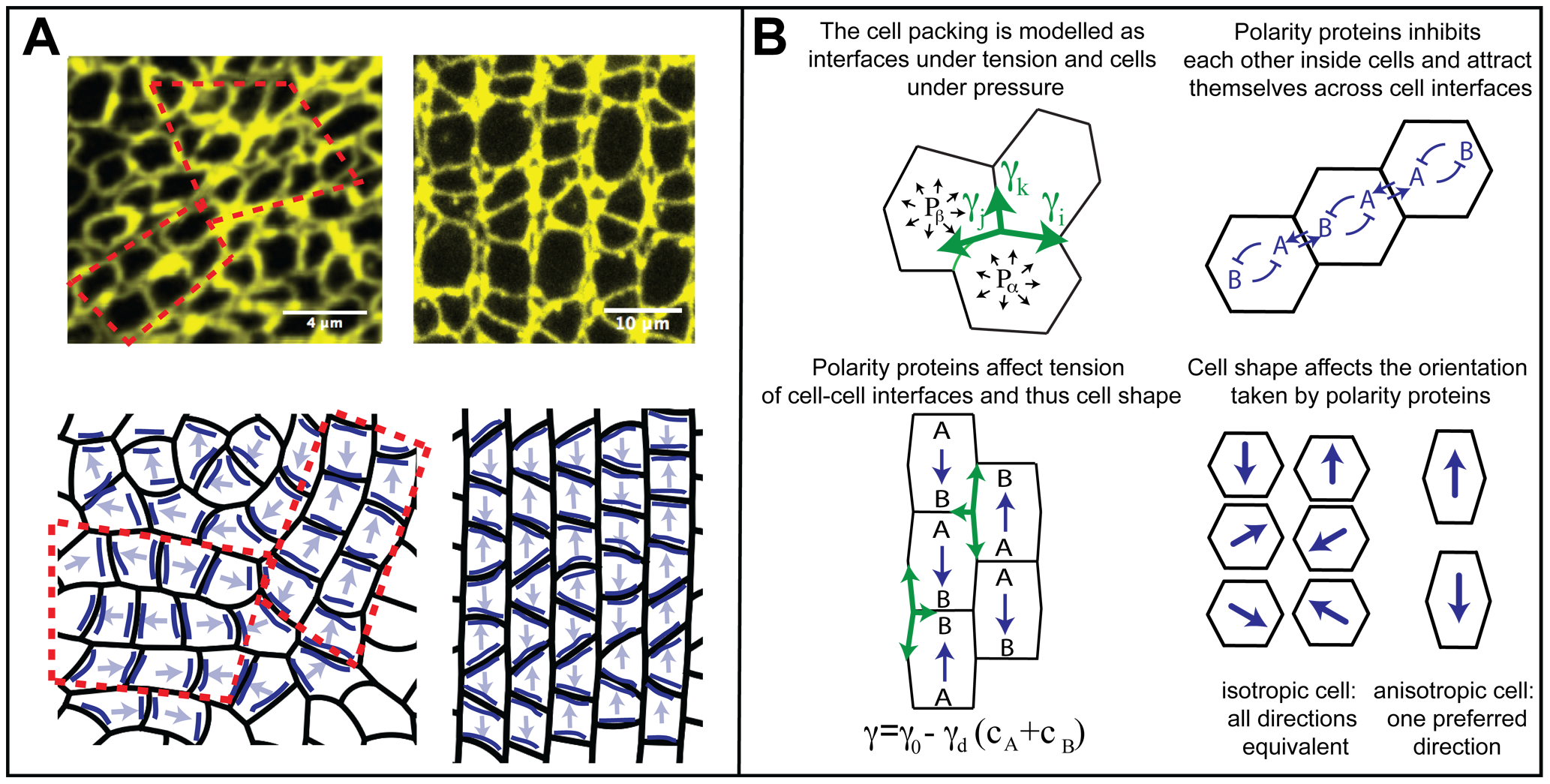
Fig. 4
Model rationale and main ingredients.
A) Cell boundaries at the level of the zonula adherens of the larval retina are revealed by ZO-1 immunostaining; note the fragments of straight, aligned rows of cones (top left, red dashed lines). We propose that this organization reflects an underlying planar cell polarity (schematic, bottom left): polarity proteins (dark blue lines) accumulate on certain interfaces, lowering their tension and leading to cell polarization (arrows). Without a global ordering signal, domains of aligned cell polarity (red dashed lines) appear. In the presence of a global ordering signal, all cones polarize in the same direction (bottom right) leading to the observed rectangular lattice and columns of cones in the adult retina (top right). B) Model ingredients: Cell shape is determined by interfacial tensions γ and pressures P; tensions must balance at vertices in mechanical equilibrium (green arrows, top left). Proteins A and B define planar cell polarity (top right) and prefer to collect on shorter interfaces (bottom right). Interfacial tensions depend on polarity protein concentrations c (bottom left).