
Image
|
Figure Caption
Fig. S2
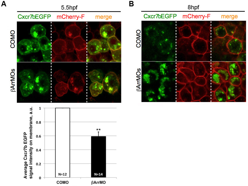
Cxcr7b localization to the plasma membrane is not enhanced by β-arrestin knockdown. Wild-type embryos were co-injected at the 1-cell stage with mRNAs for Cxcr7b-EGFP-globin32UTR and mCherry-F-globin32UTR as well as with MO mixes containing either control MO (COMO) or β-arrestin MOs. Confocal images (63×) were acquired at (A) 50% epiboly (5.5. hpf) and (B) 80% epiboly (8 hpf). The graph shows the quantification of the pixel intensity of Cxcr7b-EGFP located on the cell membrane at 5.5 hpf. The average pixel intensity in the control is set to 1. N, number of cells analyzed; **P<0.05, Student’s t-test.
Acknowledgments
This image is the copyrighted work of the attributed author or publisher, and
ZFIN has permission only to display this image to its users.
Additional permissions should be obtained from the applicable author or publisher of the image.
Full text @ Development