
|
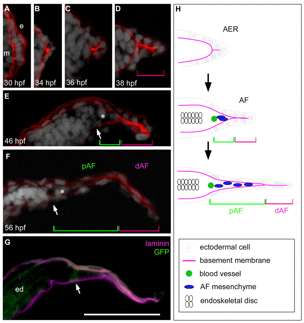
Fig. 1
AER-AF transition and morphological features in pectoral fin development. (A-G) A series of transverse pectoral fin bud sections (distal is to the right and dorsal is to the top) at the indicated stages. The basement membrane (red), shown by Laminin α5 immunostaining, is located between the ectoderm (e) and mesoderm (m) (A). Cell nuclei are visualized by DAPI (white). The distal portion of the AF (magenta brackets) consists of ectodermal cells only, and mesenchymal cells (asterisks) enter the notch of the AF within the proximal AF region (green brackets). The circumferential fin blood vessel (white arrows) is located at the base of the AF (E-G), and is recognizable by GFP distribution in fli1:EGFP y1 transgenic fish (G). ed, endoskeletal disc. Scale bar: 50 μm. (H) Diagram of transverse fin bud sections during AER/AF morphogenesis.