
|
Fig. S4
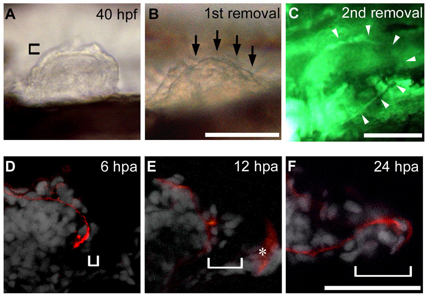
AER and AF epidermal reconstruction after AF removal. (A,B) First removal of the AF was done at 40 hpf. In this sample, the posterior two-thirds of the AF was removed (arrows in B). Note that the boundary between the remaining anterior AF and underlying endoskeletal region can be outlined, indicating that the endoskeletal region is intact after AF removal. (C) Circumferential blood vessel, which is visualized in fli1:EGFP y1 fish, after the second AF removal. As shown by white arrowheads, the entire vessel is intact. (D-F) Transverse sections immunostained for Laminin α5 (red) and DAPI (white). At 6 hours post-amputation (hpa), a layer of epidermis had regenerated (bracket in A). At 12 hpa, the AER structure had re-formed with a notch (bracket in B). Unwanted fluorescence in the trunk (asterisk) was observed in this panel. At 24 hpa, the AER had transformed into the AF structure with a long notch (bracket in C). Distal is to the right in all panels. Scale bars: 100 μm.