
|
Fig. S1
Related to Figure 2. Control of MLCK expression by microRNAs and the Dnd protein
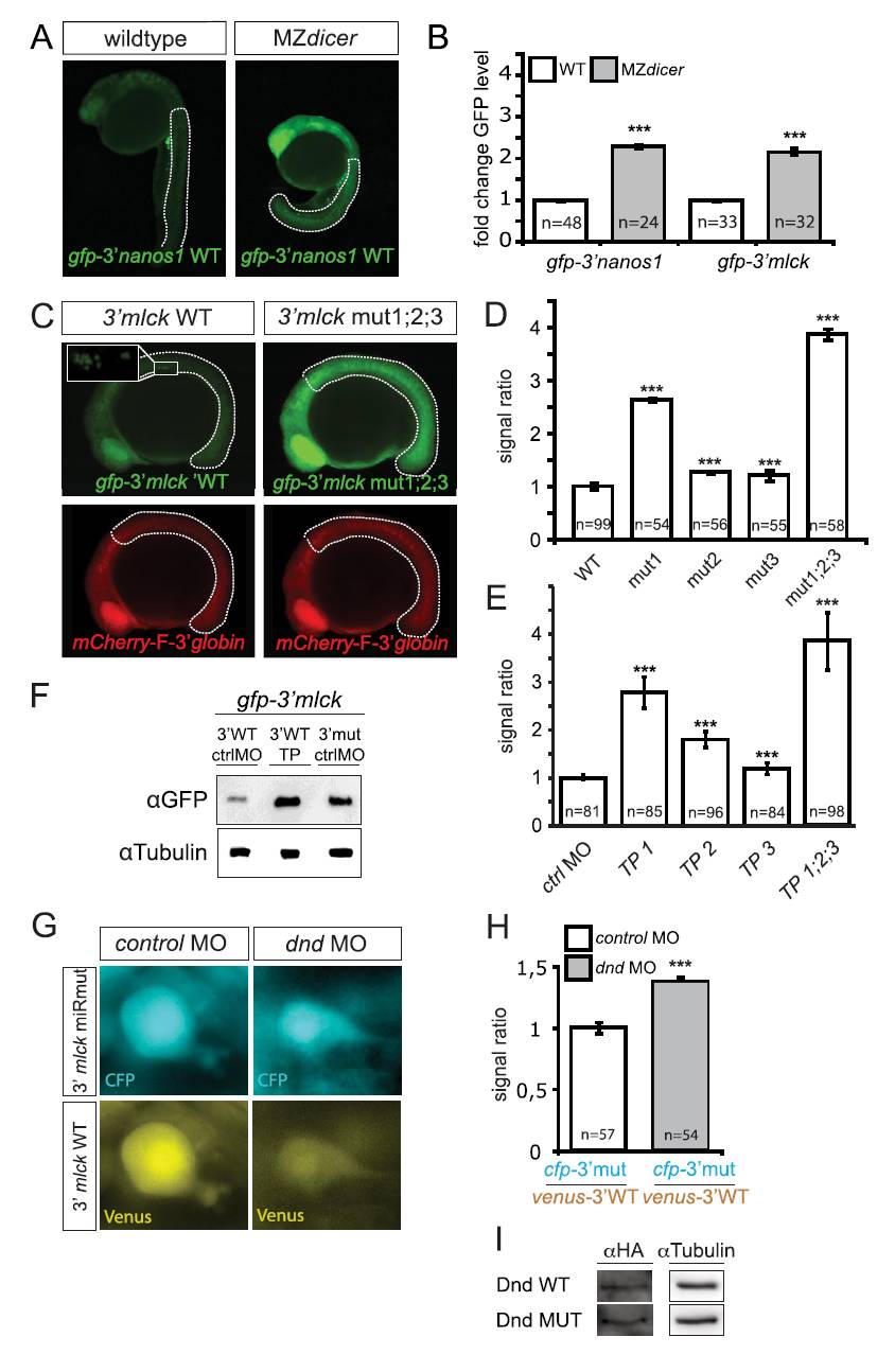
(A) Lack of miRNAs in MZdicer mutant embryos results in higher GFP expression from mRNA containing the mGFP open reading frame fused either to the 3′UTR of mlck (Figure 2A) or to the nanos 3′UTR which served as a control. Images show 24 hours post fertilization (hpf) wild-type and MZdicer embryos. (B) Quantification of the results shown in Figure 2A and Figure S1A. Dotted lines in (A) mark the measured areas; For normalization, signal intensity from co-injected mCherry-F-globin3′UTR RNA was used. (C) Inhibiting the binding of microRNAs to the 3′UTR of mlck by introducing seed-sequence mutations results in an increase in GFP expression from a reporter RNA including the miR mutations (right panel) as compared to the GFP level obtained in embryos injected with the reporter carrying the wild-type mlck 3′UTR construct (left panel). Inset shows enrichment in PGCs of GFP signal derived from the mlck 3′UTR. (D) Quantification of the results presented in panel C, showing the average GFP pixel intensity originating from the mGFP reporters containing either the wild-type mlck3′UTR or the miRNA seed mutations. Dotted lines in (C) indicate the measured areas. For normalization, signal intensity from a co-injected mCherry-F-globin3′UTR RNA was used. (E) Graphical representation of the average GFP pixel intensity originating from the mGFP reporter RNA containing the wild-type mlck 3′UTR injected together with control morpholino or with the different sets of target protectors (TPs). (F) Western blot analysis for GFP expression levels performed on embryos injected with different mlck 3′UTR reporter constructs and TPs. Application of mlck TPs or introduction of miRNA seed sequence mutations leads to increased GFP expression as compared to control. Detection of alpha-Tubulin served as control. (G) Expression of the Venus fluorescent protein from RNA containing the wild-type mlck 3′UTR is reduced in dnd MO-treated PGCs as compared to that in the RNA reporter containing the microRNA binding site mutated 3′UTR (cfp-3′mlck miRmut). (H) Normalized signal-intensity measurements in PGCs reflecting the inhibition over mlck 3′UTR in absence of Dnd function. (I) A western blot showing the input protein levels used in Figure 2I.
Reprinted from Developmental Cell, 23(1), Goudarzi, M., Banisch, T.U., Mobin, M.B., Maghelli, N., Tarbashevich, K., Strate, I., van den Berg, J., Blaser, H., Bandemer, S., Paluch, E., Bakkers, J., Toli-Nørrelykke, I.M., and Raz, E., Identification and Regulation of a Molecular Module for Bleb-Based Cell Motility, 210-218, Copyright (2012) with permission from Elsevier. Full text @ Dev. Cell