
|
Fig. 2
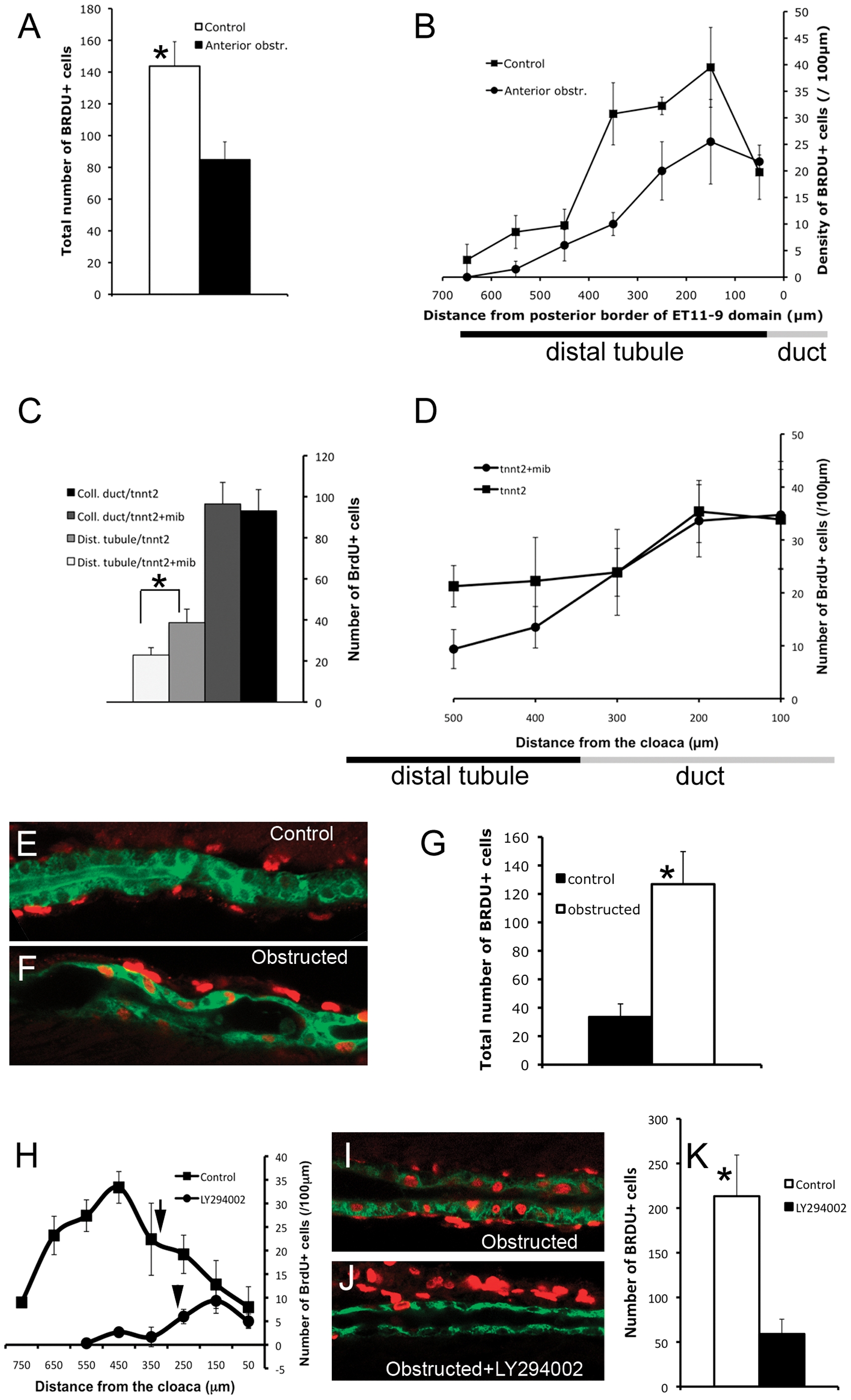
Collective epithelial migration stimulates cell proliferation in the distal tubule.
(A, B) Cell proliferation in the distal tubule (ET11-9 GFP domain) after anterior obstruction. Obstruction was induced at 30 hpf, BrdU incorporation was assessed between 2 and 3 dpf. (A) Total number of BrdU+ nuclei in the distal ET11-9 domain. White bar: control (n = 4), black bar: anterior obstruction (n = 4). P = 0.01. (B) Spatial distribution of the BrdU-positive nuclei per 100 µm length of the distal tubule (measured from the posterior border of the ET11-9 domain). Squares: control (n = 4), Circles: anterior obstruction (n = 4). (C,D) Cell proliferation in the distal nephron in mindbomb mutants. mindbomb (mib) heterozygotes were in-crossed and injected with tnnt2 morpholino to control for vascular defects in mib mutants. BrdU incorporation was assessed between 2 and 3 dpf. Homozygous mib mutants were separated from their siblings based on their axis curvature phenotype. (E) Total amount of BrdU incorporation in pronephric duct (black bar: control, n = 8; dark-grey bar: mindbomb, n = 8; P>0.05) and in the posterior distal tubule (light grey bar: control, n = 8; white bar: mindbomb, n = 8; P<0.01). (D) Linear density of the BrdU+ nuclei. Squares: tnnt2MO only control (n = 4); circles: mindbomb +tnnt2MO (n = 4). The underlying bars (B,D) indicate the position of distal tubule/pronephric duct border. (E) 24 hour BrdU incorporation in the posterior proximal tubule (2–3 dpf). (F) BrdU incorporation during 24 hour post-obstruction (2–3 dpf). (G) total number of BrdU positive nuclei in the distal 600 µm of proximal tubule. Black bar: control (n = 4), white bar: obstructed nephrons (n = 8); P<0.01. (H) Proliferation profile in control ET11-9/Tg(atp1a1a.4:GFP) transgenic fish compared to LY294002 treated fish (2–3 dpf). Arrows point to the location of distal tubule/pronephric duct interface. (I) Up-regulation of BrdU incorporation in stretched proximal tubule between 12 and 36 hours post-obstruction. (J) BrdU incorporation was markedly reduced in LY294002 treated, obstructed tubules. (K) Total number of BrdU-positive nuclei in the anterior 500 μm of the proximal tubule. White bar: BrdU incorporation in obstructed nephron/−LY294002 (n = 3), black bar: BrdU incorporation in obstructed nephron/+LY294002 (n = 3); P<0.05. (E,F) Green: GFP (ET33d10 GFP); (I,J) Green – cadherin17; (E,F,I,J) Red: BrdU.