
|
Fig. S2
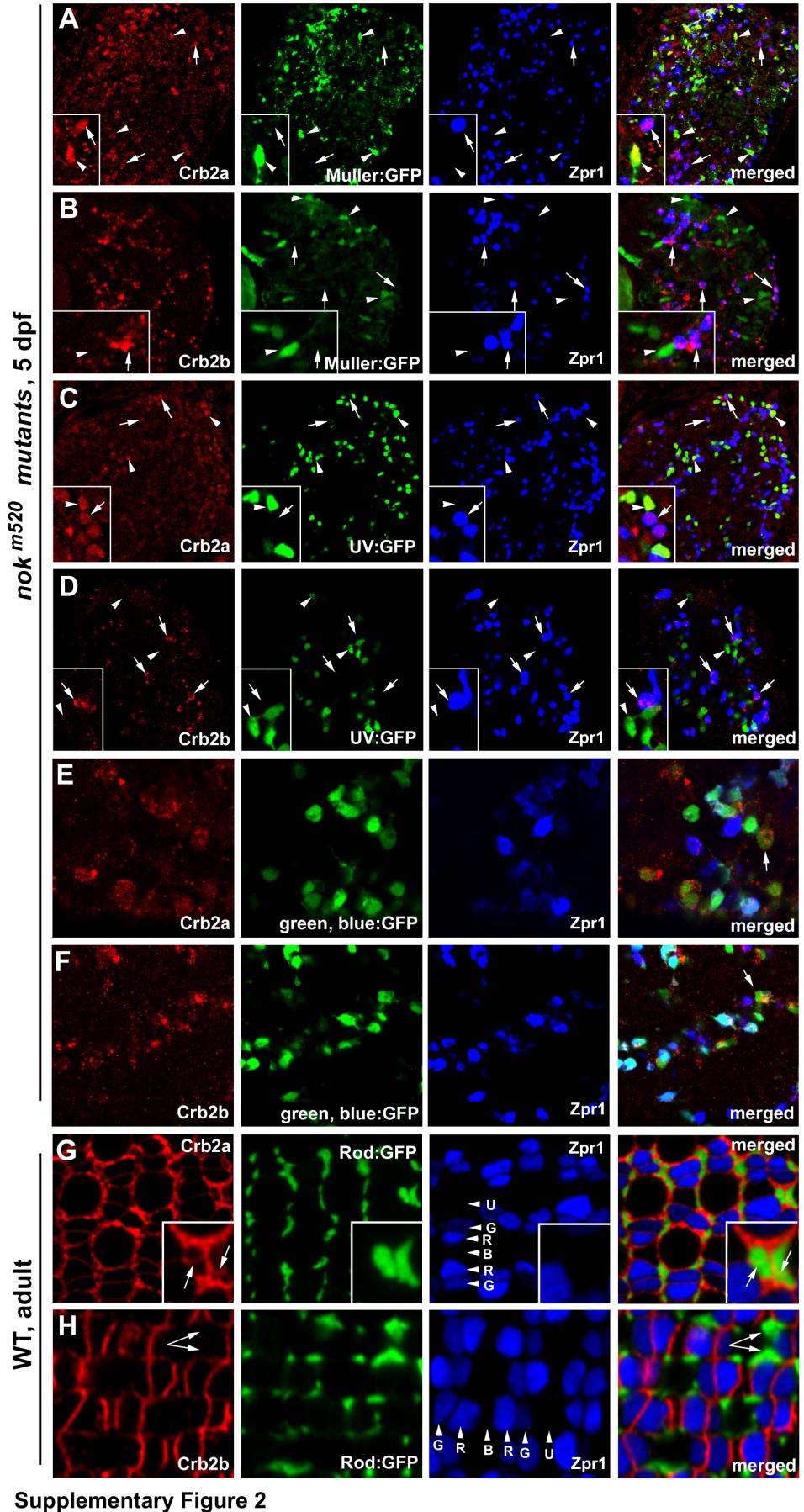
Crb2a is expressed in all types of cones, rods, and Müller cells, whereas Crb2b is predominantly expressed in green, blue and red cones but not in UV cones, rods, and Müller cells, related to Figure 2.
The distribution of Crb2a and Crb2b was examined in 5-dpf nagie oko (nok) mutant retinas (panels A-F), where retinal cells scatter irregularly and Crb proteins are internalized (Wei et al., 2006a), as well as in wildtype adult retina (panels G and H). A, B. Crb2a and Crb2b expression patterns were examined in nokm520 mutant retinas in the Tg(gfap:GFP) mi2001 transgenic background, in which Müller cells were visualized by GFP (Bernardos and Raymond, 2006). C, D. Crb2a and Crb2b expression patterns were examined in nokm520 mutant retinas in the Tg(SWS1:GFP) transgenic background, in which UV cones were visualized by GFP (Takechi et al., 2003). E, F Crb2a and Crb2b expression patterns were examined in nokm520 mutant retinas in the Tg(RH2-1:GFP) pt112 transgenic background, in which blue and green cones (arrows) expressed stronger GFP signals than red cones (Zou et al., 2010). Thus, blue cones were positive expression of GFP and negative for Zpr1. G, H. Compared to cones, the inner segment of rods, which were visualized by GFP expression in the Tg(-3.7rho:EGFP)kj2 transgenic background (Hamaoka et al., 2002), are very thin and frequently form clusters around UV cones. Crb2a expression was detected at the interfaces of the rod inner segment clusters (G, arrows). By contrast, no Crb2b signals were detectable at the rod inner segment clusters (arrows). Lettered arrowheads indicate green (G), red (R), blue (B) and UV (U) cones. Double cones were visualized with zpr1 antibodies (blue). Insets show enlarged areas.
Reprinted from Developmental Cell, 22(6), Zou, J., Wang, X., and Wei, X., Crb Apical Polarity Proteins Maintain Zebrafish Retinal Cone Mosaics via Intercellular Binding of Their Extracellular Domains, 1261-1274, Copyright (2012) with permission from Elsevier. Full text @ Dev. Cell