
|
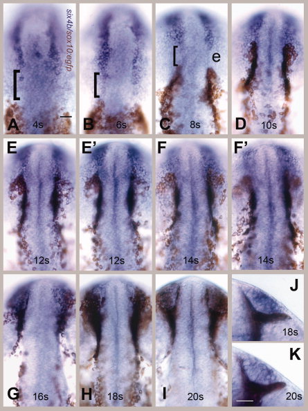
Fig. 3 six4b-expressing olfactory placode (OP) precursors do not share a common border with cranial neural crest cells (CNCCs). Visualization of OP field using six4b (in blue) and CNCCs using anti-green fluorescent protein (GFP) immunocytochemistry (in brown) in sox10:EGFP embryos. A–I: Dorsal views, rostral to the top of the page. E,E′: Two different focal planes of same embryo at 12 somites: E, dorsal OP; E′, ventral edge of the OP. F,F′: Two different focal planes at 14s (somite stage): F dorsal OP, F′ ventral edge of the OP. The neural crest cells are both dorsal and ventral to the olfactory fields at these stages. G–I: CNCCs move dorsally over the forming OP. J,K: Ventral views of the formed OP surrounded by CNCCS at 18s (J) and 20s (K), rostral to top. Scale bars = 30 μm in A (applies in A–K). Twenty embryos were examined per time point.