
Image
|
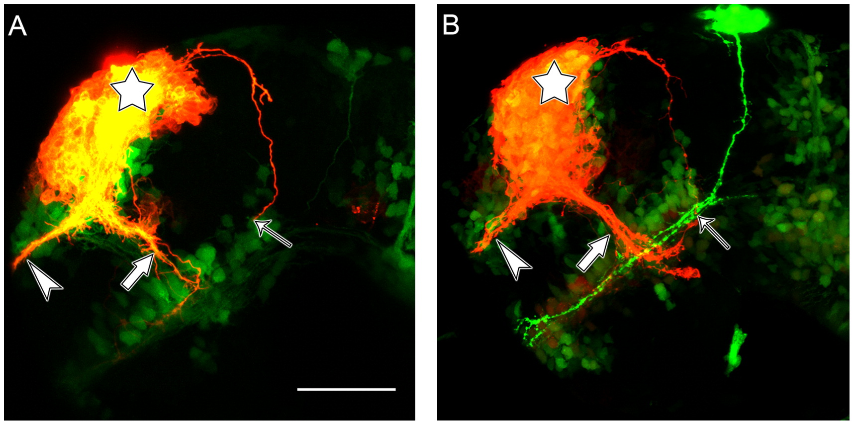
Figure Caption
Fig. 7 Tracing the EDAB to the ventrorostral diencephalic cluster. (A) HuC:kaede brain. (B) HuC:kaede/Foxd3:GFP brain. DiI labelling is represented in red and the kaede/GFP fluorescence in green. The star indicates the site of DiI injection, the arrowhead indicates the anterior commissure, the large arrow is the supraoptic tract and the thin arrow points at the identifiable end of the EDAB. In (B) the DVDT trajectory is clearly depicted by GFP fluorescence in green. Scale bar=50 μm.
Acknowledgments
This image is the copyrighted work of the attributed author or publisher, and
ZFIN has permission only to display this image to its users.
Additional permissions should be obtained from the applicable author or publisher of the image.
Reprinted from Developmental Biology, 367(2), Gaudin, A., Hofmeister, W., and Key, B., Chemoattractant axon guidance cues regulate de novo axon trajectories in the embryonic forebrain of zebrafish, 126-139, Copyright (2012) with permission from Elsevier. Full text @ Dev. Biol.