
|
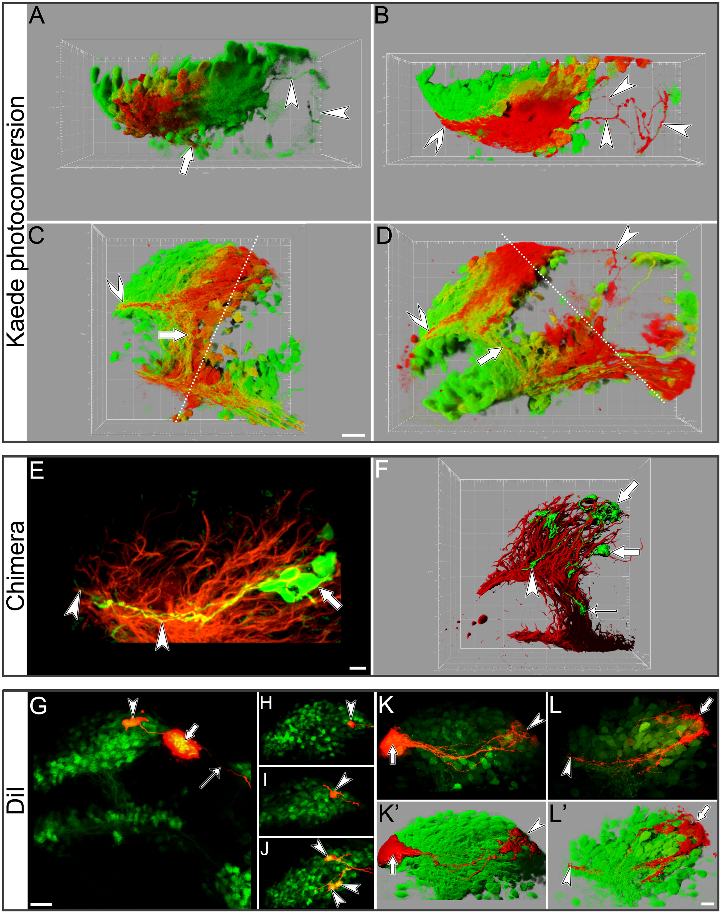
Fig. 6 EDAB originates from a subset of neurons in the caudal portion of the DRC that normally project into the anterior commissure. Axon tracing using kaede photoconversion (A–D), chimeras (E–F) and DiI (G–L′). All images are confocal image stacks processed with Imaris software. The resulting representation are either a simple maximum intensity projection under orthogonal perspective along the Z axis (E–K), perspective views of shadow projection rendering of the stacks (A–D, K′, L′) or a mix of shadow projection rendering and volume rendering (F). (A) Dorsal view of the DRC in a dcc knock down embryo at 30 hpf. When the anterior portion of the DRC was photoconverted (red), the EDAB remained green (arrowheads) while red photoconverted axons exited via the SOT (large arrow). (B) Dorsal view of the DRC in a dcc knock down embryo at 30 hpf. When the posterior portion of the DRC was photoconverted (red) the EDAB was red (arrowheads). Note also the large number of photoconverted axons extending towards the anterior commissure (chevron). (C) Lateral view of a control brain at 30 hpf. The dotted line indicates the path of the photoconverting laser through the DRC and the underlying VRC. When the posterior portion of the DRC is photoconverted (red) in a control brain, both the anterior commissure (chevron arrow) and SOT (large arrow) are labelled in red. (D) Lateral view of a dcc knock down embryo at 30 hpf. The dotted line represents the path of the photoconverting laser through the DRC and the caudal VRC and TPOC. The EDAB (arrowhead), anterior commissure axons (chevron arrow) and SOT axons (large arrow) are all labelled in red. (E) Lateral view of the DRC in a wild-type host chimeric animal immunolabelled for expression of acetylated α-tubulin (red) showing the axon trajectory of donor neurons expressing kaede from the caudal edge of the DRC (large arrows) into the anterior commissure (arrowheads). (F) Lateral view of the forebrain of a wild-type host chimeric animal immunolabelled for expression of actetylated α-tubulin (red) showing growth cones of kaede-expressing donor cells (large arrow) in the anterior commissure tract (arrowhead) and the SOT (thin arrow), representing the two routes used by axons originating from the caudal half of the DRC. (G) Lateral view of the forebrain of a dcc knock down embryo showing the site of microiontophoretic deposit of DiI in the route of the EDAB (large arrow). The EDAB axon is anterogradely labelled (thin arrow). Retrograde labelling reveals the cell body of the neuron located at the dorsocaudal end of the DRC (arrowhead). (H)–(J) Lateral views showing the localisation of the cell bodies giving rise to the EDAB in the caudal half of the DRC (arrowheads) after retrograde DiI labelling of the EDAB. (K)–(K)′ Lateral view of the DRC in a control embryo after injection of DiI in the anterior commissure (large arrow). Axons are retrogradely labelling the cell bodies located at the dorsocaudal end of the DRC (arrowhead). (L)–(L)′ Lateral view of the DRC in a control embryo after injection of the DiI at the dorsocaudal end of the DRC (large arrow) labelling axons anterogradely in the tract of the anterior commissure. These axons enter the anterior commissure after passing the point where the SOT bifurcates from the tract. Scale bars=15 μm (A–D), 5 μm (E), 10 μm (F), 20 μm (G), 25 μm (H–J), 10 μm (K–L′).
Reprinted from Developmental Biology, 367(2), Gaudin, A., Hofmeister, W., and Key, B., Chemoattractant axon guidance cues regulate de novo axon trajectories in the embryonic forebrain of zebrafish, 126-139, Copyright (2012) with permission from Elsevier. Full text @ Dev. Biol.