
Fig. 6
- ID
- ZDB-IMAGE-120612-6
- Publication
- Moro et al., 2012 - In vivo Wnt signaling tracing through a transgenic biosensor fish reveals novel activity domains
- All Figures
- Figures for Moro et al., 2012
|
Fig. 6
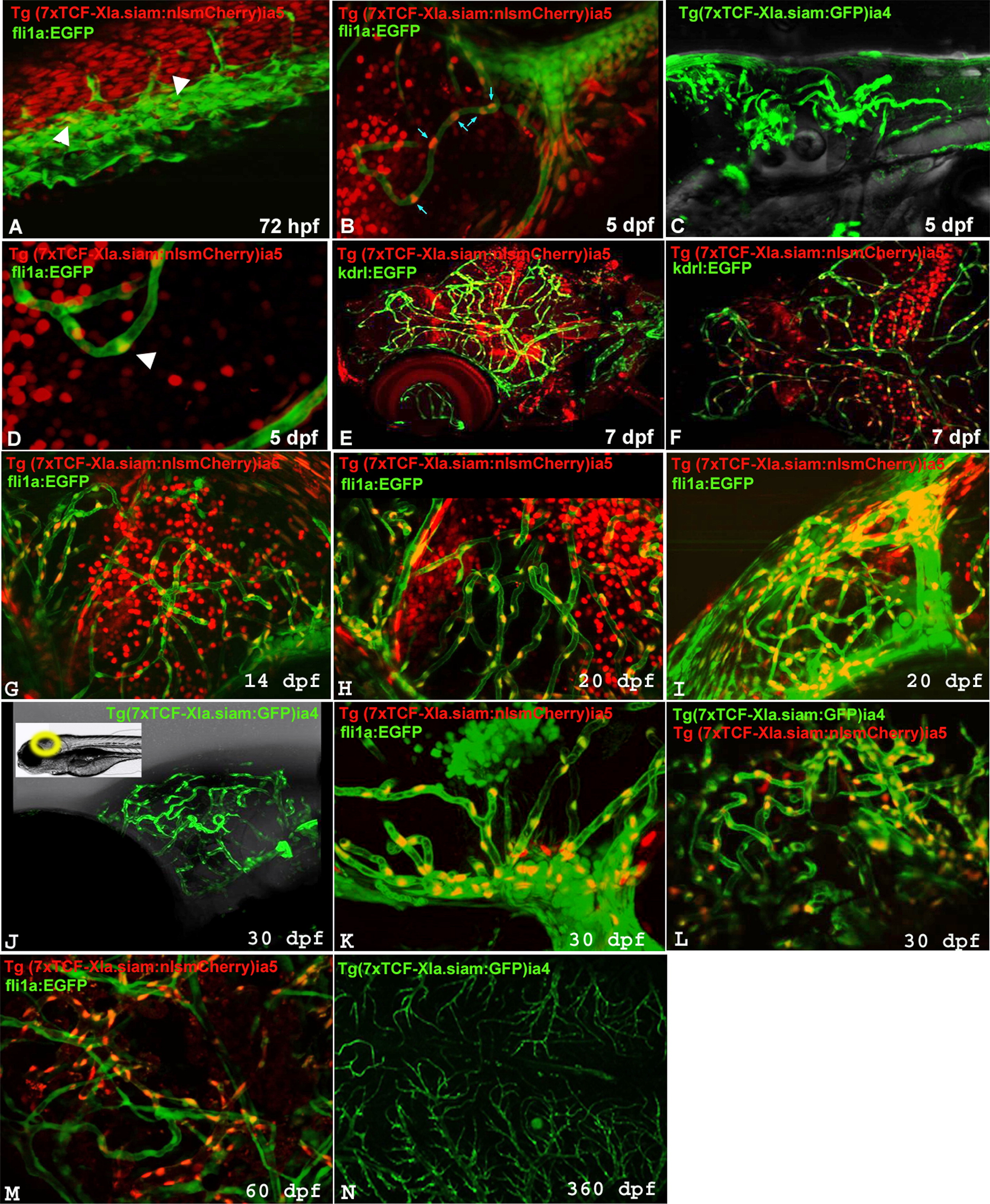
TCFsiam transgene expression in the CNS endothelial compartment during larval, juvenile and adult stages. (A) Confocal image of the trunk vessels in a representative 72 hpf Tg(fli1a:EGFP)y1/ Tg(7xTCF-Xla.Siam:nlsmCherry)ia5 fish. Few endothelial cells are positive for the transgene (arrowheads in A). (B) Confocal image of the CNS vessels in a 5 dpf representative Tg(fli1a:EGFP)y1/Tg(7xTCF-Xla.Siam:nlsmCherry)ia5 fish. Several endothelial fli1a:EGP expressing cells are positive for the Tg(7xTCF-Xla.Siam:nlsmCherry)ia5 transgene (highlighted by light blue arrows). (C) Confocal Z-stack projection of a 5 dpf Tg(7xTCF-Xla.Siam:GFP)ia4, showing the targeted labeling of the cerebral vascular network. (D) Magnification of a CNS blood vessel of a representative 5 dpf Tg(fli1a:EGFP)y1/Tg(7xTCF-Xla.Siam:nlsmCherry)ia5 larva. A double positive endothelial cell is marked by a arrowhead. (E,F) Confocal Z-stack projection of a representative 7 dpf Tg(kdrl:EGFP)s843/Tg(7xTCF-Xla.Siam:nlsmCherry)ia5, showing a perfect localization of the TCFsiam signal in the CNS endothelial compartment. In F a higher magnification of (E) is shown. (G) Confocal Z-stack projection in the midbrain area of a representative 14 dpf Tg(fli1a:EGFP)y1/Tg(7xTCF-Xla.Siam:nlsmCherry)ia5 fish. (H,I) Confocal Z-stack projection of a representative 20 dpf Tg(fli1a:EGFP)y1/Tg(7xTCF-Xla.Siam:nlsmCherry)ia5 fish in the midbrain area (H) and in the forebrain area (I), showing strong labeling of the brain vessels in the transgenic reporter fish. (J) Confocal projection a 30 dpf fish brain showing localized expression of the Tg(7xTCF-Xla.Siam:GFP)ia4 transgene in the vascular compartment of the brain. A small inset illustrates the area analyzed by confocal microscopy in the living fish. (K, L, M) Confocal Z-stack projection of a representative 30 dpf (K) and 60 dpf (M) Tg(fli1a:EGFP)y1/Tg(7xTCF-Xla.Siam:nlsmCherry)ia5 and 30 dpf (L) Tg(7xTCF-Xla.Siam:GFP)ia4/Tg(7xTCF-Xla.Siam:nlsmCherry)ia5 fish showing a persistent expression of the TCFsiam transgene in the brain vascular network. (N) Confocal acquisition of a whole fixed brain from a one year old fish demonstrating long-term expression of the Tg(7xTCF-Xla.Siam:GFP)ia4 transgene in CNS vascular network.
Reprinted from Developmental Biology, 366(2), Moro, E., Özhan, G., Mongera, A., Beis, D., Wierzbicki, C., Young, R.M., Bournele, D., Domenichini, A., Valdivia, L.E., Lum, L., Chen, C., Amatruda, J.F., Tiso, N., Weidinger, G., and Argenton, F., In vivo Wnt signaling tracing through a transgenic biosensor fish reveals novel activity domains, 327-340, Copyright (2012) with permission from Elsevier. Full text @ Dev. Biol.