
|
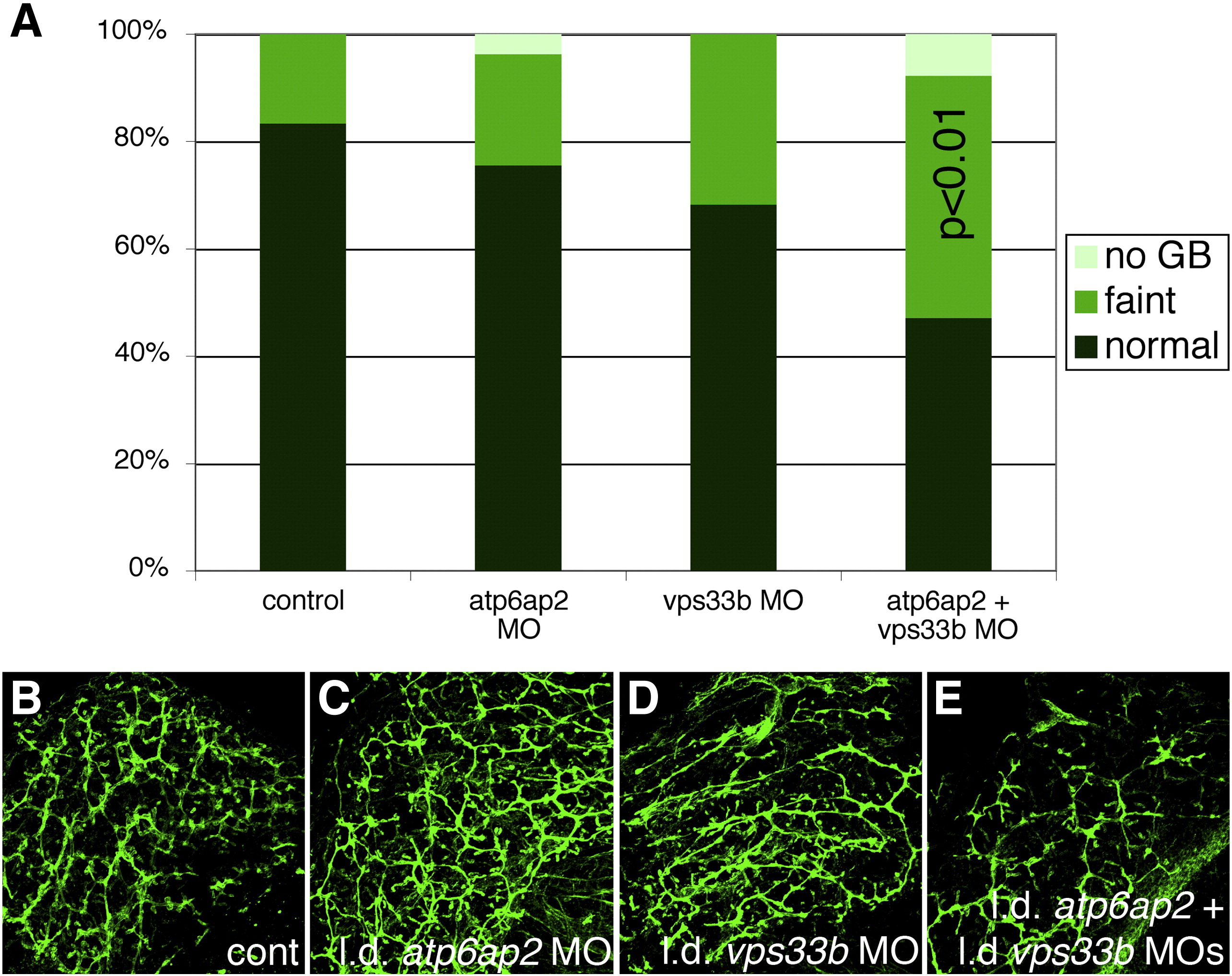
Fig. 7
Additive effect of vps33b and atp6ap2 inhibition. (A) Gallbladder PED-6 uptake in 5 dpf control larvae (cont), larvae injected with a low dose of atp6ap2 morpholino (MO), larvae injected with a low dose of vps33b MO, and larvae injected with a combination of the low dose MOs. The number of larvae with faint gallbladders was not significantly different between either of the MOs alone compared to control, but the number of larvae with faint gallbladders after injection of the combination of MOs was significantly increased compared to control (p < 0.01) and to either of the MOs alone (p < 0.01 for atp6ap2, p < 0.05 for vps33b). (B–E) Confocal projections of cytokeratin immunostaining from 5 dpf larvae corresponding to the conditions depicted in the graph in (B), showing control (B, cont), low-dose atp6ap2 MO (C), low-dose vps33b MO (D), and the combination of the two MOs in low dose (E). Note that (C) and (D) are similar to (B), while (E) more closely resembles the pn defects shown above. Samples depicted in B–E are representative of pooled samples of all PED-6 classifications.
Reprinted from Developmental Biology, 365(2), Eauclaire, S.F., Cui, S., Ma, L., Matous, J., Marlow, F.L., Gupta, T., Burgess, H.A., Abrams, E.W., Kapp, L.D., Granato, M., Mullins, M.C., and Matthews, R.P., Mutations in vacuolar H(+)-ATPase subunits lead to biliary developmental defects in zebrafish, 434-444, Copyright (2012) with permission from Elsevier. Full text @ Dev. Biol.