
|
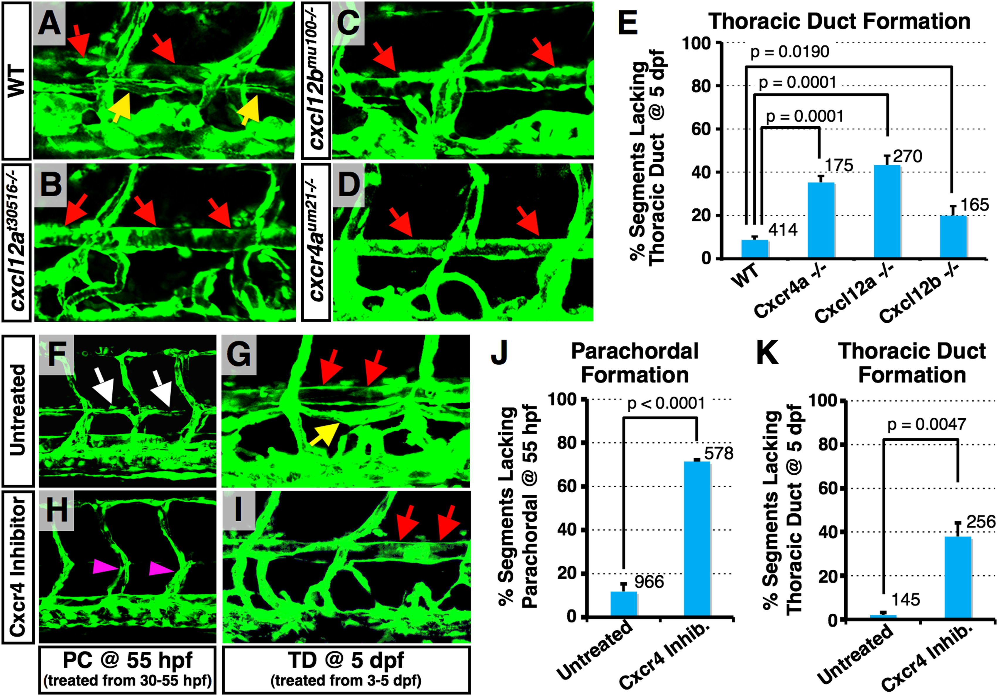
Fig. 5
Deficiency of Chemokine Signaling Results in the Loss of Lymphatic Network (A–D) Confocal images of trunk vessels in 5 dpf Tg(fli-EGFP)y1 wild-type (A), cxcl12at30516/ (B), cxcl12bmu100/ (C), and cxcr4aum21/ (D) homozygous mutant animals. Red and yellow arrows indicate DA and TD, respectively. (E) Quantification of TD formation defect in 5 dpf mutant animals. Values are the mean ± SEM. Numbers of counted TD segments are indicated above the bars. (F–I) Confocal images of trunk vessels in 55 hpf Tg(fli-EGFP)y1 animals treated from 30 to 55 hpf (F) and (H) or 5 dpf Tg(fli-EGFP)y1 animals treated from 3 to 5 dpf (G) and (I) with either DMSO vehicle (F) and (G) or Cxcr4 inhibitor (H) and (I). (J) Quantification of the PC formation defect in 55 hpf DMSO vehicle or Cxcr4 inhibitor-treated embryos. Values are the mean ± SEM. (K) Quantification of the TD formation defect in 5 dpf DMSO vehicle or Cxcr4 inhibitor-treated embryos. Values are the mean ± SEM. In (J) and (K), numbers of counted PC or TD segments are indicated above the bars. See also Figure S5.
Reprinted from Developmental Cell, 22(4), Cha, Y.R., Fujita, M., Butler, M., Isogai, S., Kochhan, E., Siekmann, A.F., and Weinstein, B.M., Chemokine Signaling Directs Trunk Lymphatic Network Formation along the Preexisting Blood Vasculature, 824-836, Copyright (2012) with permission from Elsevier. Full text @ Dev. Cell