
|
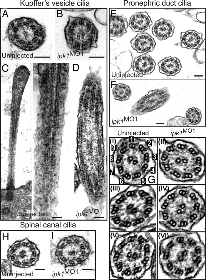
Fig. 2
ipk1 knockdown does not affect ciliary axoneme organization. (A and B) TEM cross-sections of cilia in KV for uninjected (A) and ipk1 MO1 (B) embryos. (C and D) Longitudinal TEM sections showing axonemal microtubules in KV cilia in uninjected (C) and ipk1 MO1 (D) embryos. (E–I) TEM sections showing pronephric duct cilia (E, F, and G) and spinal canal cilia (H and I). Enlarged images of independent pronephric duct cilia TEM sections are shown in G. Cilia sections in uninjected (E, G-I, G-III, G-V, and H) and ipk1 MO1 (F, G-II, G-IV, G-VI, and I) embryos are shown. In G–I, numerical tags are assigned to each of the nine outer microtubule doublets for reference to dynein arms in the text. (Scale bars, 0.1 μm.)