
|
Fig. S4
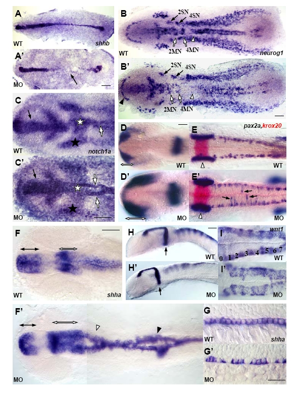
NBP is required for embryo development. All paired panels compare NBP MO morphants (MO) with wild-type embryos (WT). A-F′, I, I′ show flat preparations, anterior to the left. G-H′ show lateral view, anterior to the left. (A,A′) shhb (two-somite stage). Arrow in A′ indicates the area in which cells of the presumptive floor plate are not specified. (B,B’) neurog1 (four-somite stage). Filled arrowhead indicates the reduced telencephalon in NBP MO-injected embryo, open arrows point to motoneurons of rhombomeres 2 and 4 (2MN and 4MN), filled arrows point to sensory neurons of rhombomeres 2 and 4 (2SN and 4SN), open arrowheads point to the ventral neural clusters, not clearly distinguishable in the morphants. (C,C′) notch1a (tailbud/one-somite stage, head region). Filled arrow indicates the area of expression in the presumptive forebrain broader and shorter in the NBP MO-injected embryo, filled stars indicate the lateral stripes of expression in the ectoderm, open stars point to the area of high transcript density in the prospective hindbrain, almost missing in the morphant, open arrows indicate the midline stripe of expression in the prechordal mesoderm splitted in the NBP MO-injected embryo. (D-E′) pax2a, krox20 (18-somite stage, D,D′: head region; E,E′: hindbrain region). Open arrows indicate the length of pax2a expression in the optic primordial, arrows indicate scattered interneurons in hindbrain area, open arrowheads indicate the otic vesicle. (F-G′) shha (21-somite stage; F,F′: head-trunk region; G,G′: trunk region, note the stained floorplate cells). Filled arrows indicate the ventral anterior diencephalon reduced in the morphant, open arrows point to the ventral midbrain expanded in the NBP morphant, open arrowhead indicates the splitted expression in the hindbrain, filled arrowhead indicates the split in the spinal cord. (H-I′) wnt1 (25-somite stage; H,H’: head-trunk region; I,I′: hindbrain region). Filled arrows indicate the midbrain hindbrain boundary, numbers mark locations of the corresponding rhombomeres. Scale bar =50 μm.
Reprinted from Developmental Biology, 365(1), Boggetti, B., Jasik, J., Takamiya, M., Strähle, U., Reugels, A.M., and Campos-Ortega, J.A., NBP, a zebrafish homolog of human Kank3, is a novel Numb interactor essential for epidermal integrity and neurulation, 164-174, Copyright (2012) with permission from Elsevier. Full text @ Dev. Biol.