
|
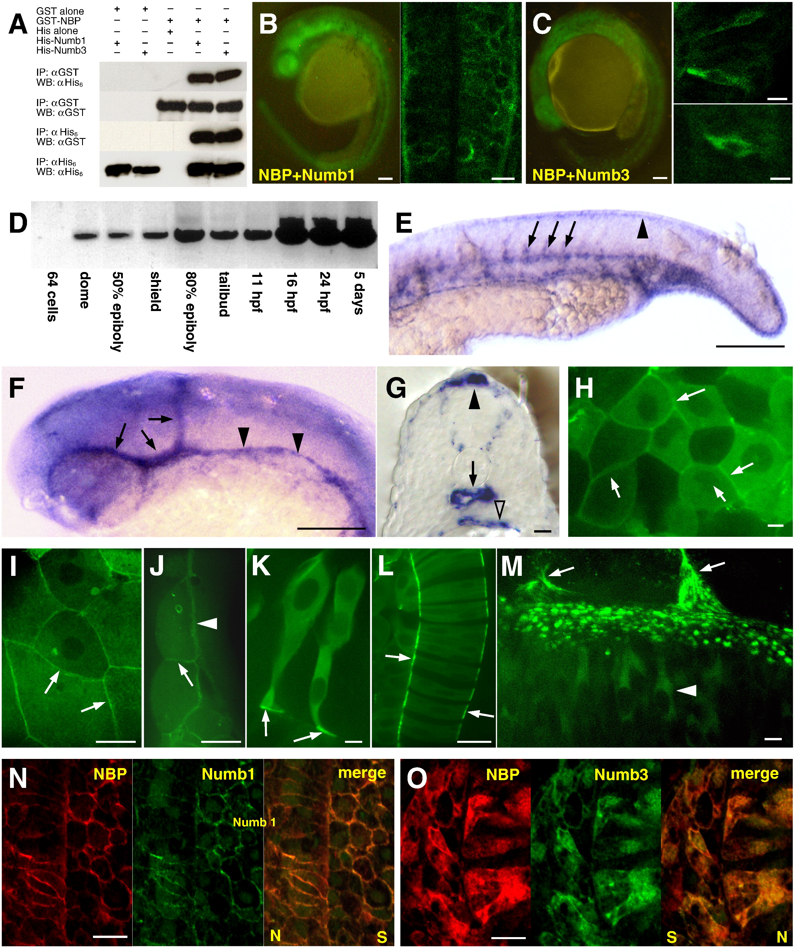
Fig. 2
NBP and Numbs interact in vitro and in vivo. (A) Immobilized GST or GST–NBP fusion protein were mixed with either His alone, His-Numb1 or His-Numb3. The complexes were washed, analyzed by SDS-PAGE, and immunoblotted with either anti-His6 (top panel) or anti-GST (second panel). Immobilized His, His-Numb1 or His-Numb3 fusion proteins were mixed with either GST alone or GST–NBP. The complexes were washed, analyzed by SDS-PAGE and immunoblotted with either anti-GST (third panel) or anti-His6 (bottom panel). (IP: immunoprecipitation; WB: Western blot). (B) YFP in the BiFC assay was restored due to the NBP–Numb1 interaction in the whole embryo (left) and in the cells the fluorescent signal was detected at their periphery (right picture is showing cells of the neural tube). (C) NBP–Numb3 association resulted in the formation of YFP bimolecular complex in the whole embryo (left). At the cellular level, YFP signal was localized in the cytoplasm (right, upper picture represents the retina, bottom picture a cell of the neural tube). NBP exhibits distinct expression and localization patterns. (D) RT-PCR results showed that NBP expression started at dome stage and progressively increased during development. (E–G) Distribution of NBP transcripts revealed by whole-mount in situ hybridization. 24 hpf embryos (E, F lateral view, anterior to the left; G, transversal section of trunk region). Transcripts of NBP became concentrated in vascular endothelial cells of the head (arrows in E), axial (arrowheads in E) and intersomitic vessels (arrows in F), and in the median fin fold (arrowhead in F). Section in G shows NBP expression in the epidermis, median fin fold (filled arrowhead), at the basal pole of neural tube cells, in the dorsal aorta (arrow) and in the gut (open arrowhead). (H–M) NBP–GFP fusion protein localization. During gastrulation (ca. 75% epiboly; H–J) NBP–GFP is distributed uniformly in the cytoplasm. In deep cells it accumulates at the periphery in correspondence with the cell–cell contacts (arrows in H). In the EVL (I, J), the signal intensity significantly increases at the basal and lateral membranes (J, arrowhead points to the contact between EVL and yolk cell, arrow points to the contact between neighboring EVL cells; I shows a tangential section through the EVL covering deep cells, arrows point to lateral membranes). In two- to four-somite stage embryos, the cells exhibit cytoplasmic localization (K, L). In neuronal cells (K), NBP is accumulated at the basal pole at the contact with the basement membrane (arrows) and in notochord cells at the notochordal sheath (arrows in L). In 24 hpf embryos, NBP is cytoplasmic in neural tube cells (arrowhead in M points to a single neuronal cell). Z projection shows the punctuated pattern of neuronal cells-basement membrane attachments (arrows point to somitic boundaries). (N, O) Immunodetection of NBP and Numbs. For proteins co-localization, the antibody against c-myc (for NBP) was growing also in mouse but detected with a secondary antibody coupled with cy3, the antibody against HA (for Numbs) was growing in rat and detected by a secondary antibody conjugated with FITC. NBP and Numb1 when co-expressed were localized exclusively at the cell periphery (N). When NBP was co-expressed with Numb3, both proteins were distributed through the whole cells (O). N and O show the area around the border between the neural tube (N) and the somite (S). Scale bar: B and C left panels, E, F = 100 μm, B and C right panels, G–J, L, N, O = 10 μm, M = 5 μm, and K = 2 μm.
Reprinted from Developmental Biology, 365(1), Boggetti, B., Jasik, J., Takamiya, M., Strähle, U., Reugels, A.M., and Campos-Ortega, J.A., NBP, a zebrafish homolog of human Kank3, is a novel Numb interactor essential for epidermal integrity and neurulation, 164-174, Copyright (2012) with permission from Elsevier. Full text @ Dev. Biol.