
|
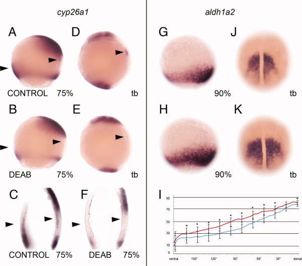
Fig. 3
A–K: Perturbation of cyp26a1 (A–F) and aldh1a2 (G–K) expression domains in the gastrula hypoblast upon 10-5M diethylaminobenzaldehyde (DEAB) treatments from 30% epiboly until the stage of fixation. Animal is to the top and dorsal to the right (A,B,D,E,G,H) or shown in dorsal view (J,K) and in parasagittal sections (C,F). A–F: DEAB treatments lead to an absence of the paraxial cyp26a1 domain at 75% and to a notable reduction of the marginal hypoblast expression domain (compare expression domains in A,D,C to B,E,F, arrowheads). G,H,J,K: DEAB treatments from 30% epiboly on reveal an enhanced signal intensity of the aldh1a2 expression domain ventrolaterally at 90% epiboly and tail bud stage (compare G,J to H,K). I: The graph shows the relative enhancement of marginal aldh1a2 expression at 90% epiboly of 10 experimental embryos (red) compared to 10 control embryos (blue) in one experiment; asterisks show significant differences in staining in controls vs. experimental embryos.