
Fig. S1
- ID
- ZDB-IMAGE-120405-13
- Publication
- Cadwalader et al., 2012 - 2-O-sulfotransferase regulates Wnt signaling, cell adhesion and cell cycle during zebrafish epiboly
- All Figures
- Figures for Cadwalader et al., 2012
|
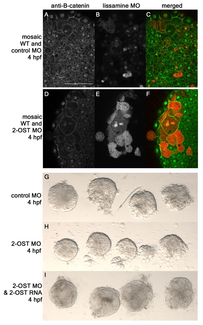
Fig. S1
Adhesion defects in 2-OST MO are cell autonomous. (A-F) Single planes of z-stack at 4 hpf of mosaic embryos formed from injecting either control MO+lissamine-conjugated MO (B,C) or 2-OST MO+lissamine-conjugated MO (E,F) into a single cell at the 32-cell stage. Anti-β-catenin for control MO mosaic (A) or 2-OST MO (D). (G-I) Animal caps removed and immediately imaged from control MO (G), 2-OST MO (H), or 2-OST MO+2-OST RNA (I) embryos to examine adhesion defects. Minimal cells were observed around control MO and rescue-injected animal caps, but many cells were observed around 2-OST MO, indicating decreased adhesion.