
|
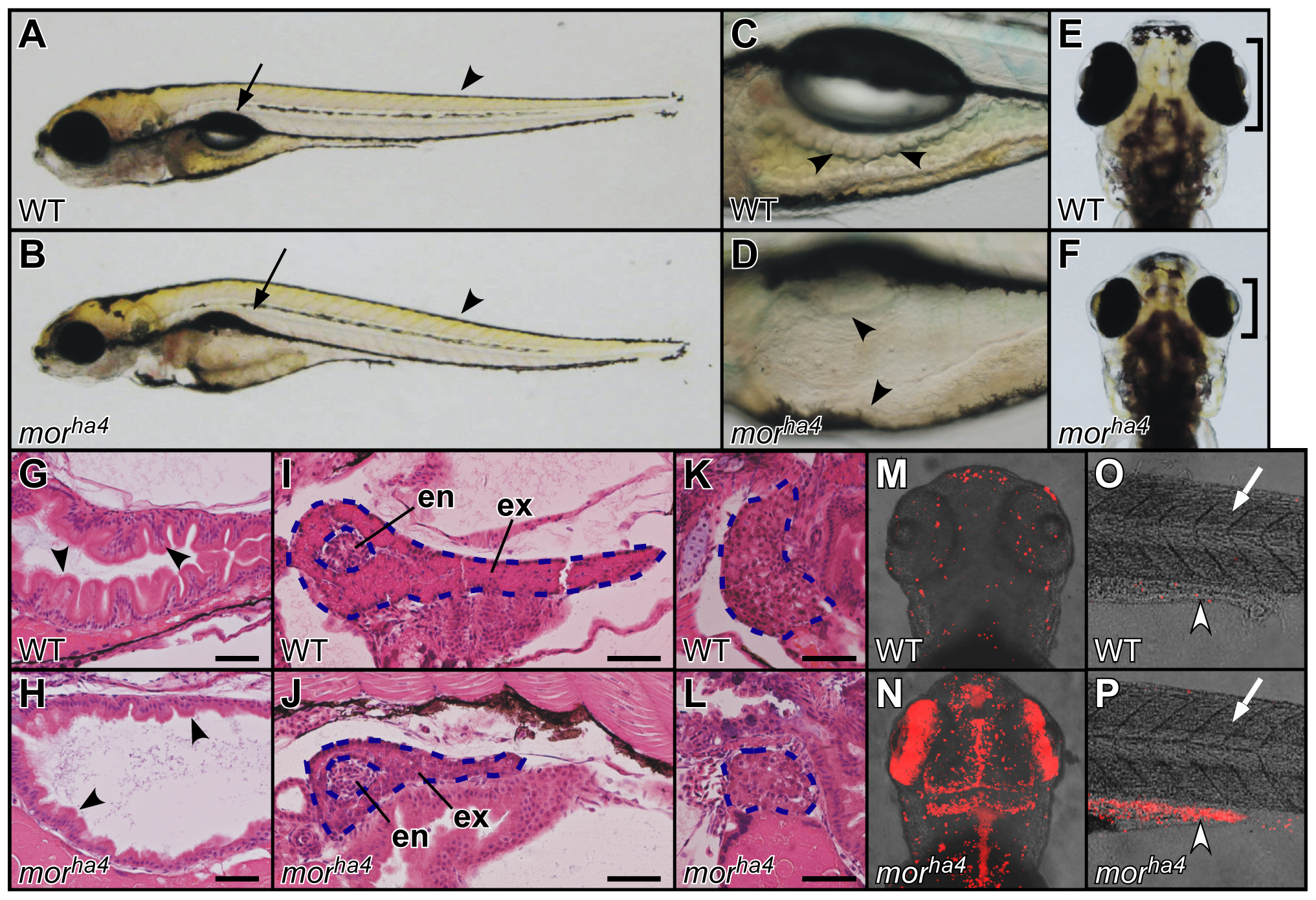
Fig. 1
Phenotype of the morha4 mutant.
(A–F) Lateral (A–D) and dorsal (E, F) views of live WT and morha4 larvae at 5.5 dpf. The swim bladder failed to inflate (arrows in A, B), the intestine lacked folds (arrowheads in C, D), and the retinae were reduced in size (brackets in E, F) in the morha4 mutant. Conversely, somite formation in the morha4 mutant appeared normal (arrowheads in A, B). (G–L) Sagittal sections of 5.5-dpf larvae stained with hematoxylin and eosin. The intestine lacked folds and was thin walled (arrowheads in G, H), and the exocrine pancreas (blue dotted lines in I, J) and liver (blue dotted lines in K, L) were small in the morha4 mutant. In contrast, the endocrine pancreas (blue dotted lines in I, J) in WT larvae was indistinguishable from that in morha4 larvae. Scale bars, 50 μm. (M–P) Dorsal views, anterior to the top (M, N). Lateral views, anterior to the left (O, P). Apoptotic cells were detected using the TUNEL method. An increase in apoptotic cells was evident in the brain, retinae, and posterior intestine of the morha4 larvae (white arrowheads in O, P) compared to WT larvae, but not in the morha4 somite (white arrows in O, P). en, endocrine pancreas; ex, exocrine pancreas.