
|
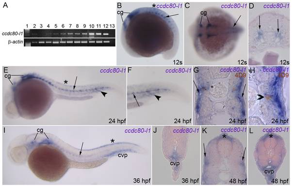
Fig. 2 Expression of ccdc80-l1 analyzed by RT-PCR and WISH.
(A) RT-PCR performed on different embryonic stages and adult tissues; the expression of ccdc80-l1 and β-actin are shown. Lanes are: ladder (lane 1), ovary (lane 2), 2–4 cells stage (lane 3), 64–1000 cells stage (lane 4), 30% epiboly (lane 5), 60–70% epiboly (lane 6), somitogenesis (lane 7), 24 hpf (lane 8), 30 hpf (lane 9), 48 hpf (lane 10), 72 hpf (lane 11), adult muscle (lane 12) and negative control (lane 13) in the absence of cDNA. (B–J) WISH performed on zebrafish embryos at several stage of development. (B, C) During somitogenesis ccdc80-l1 was expressed by cranial ganglia (cg), dorsal dermis (asterisk), adaxial cells and muscle pioneers at the level of the horizontal myoseptum (arrow). (D) ccdc80-l1 expression in a transverse section of the trunk of an embryo at 12 somites stage (arrows). (E–H) At 24 hpf, the hybridization signal was detectable in cranial ganglia (cg), dermis (asterisk), adaxial cells (arrow) and ventral somites (arrowhead). (F) Higher magnification of the tail at 24 hpf. (G) Transversal section of an embryo at 24 hpf. (H) Transversal section showing that at 24 hpf ccdc80-l1 hydridization signal co-localized with the nuclear labeling of 4D9 antibody, corresponding to the engrailed-positive muscle pioneers population (open arrowhead). (I, J) At 36 hpf, the signal of ccdc80-l1 probe was detected in cranial ganglia (cg), migrated adaxial cells (arrow), dorsal dermis (asterisk) and caudal vein plexus region (cvp). (K, L) At 48 hpf, ccdc80-l1 was detected in dorsal dermis (asterisk), external adaxial cells (arrows in K) and caudal vein plexus region (cvp in L). (B, E, F, I) Lateral views; dorsal is up, anterior is left; (C) dorsal view, anterior is left; (D,G, H, J–L) transversal sections, dorsal is up.