
Image
|
Figure Caption
Fig. 2
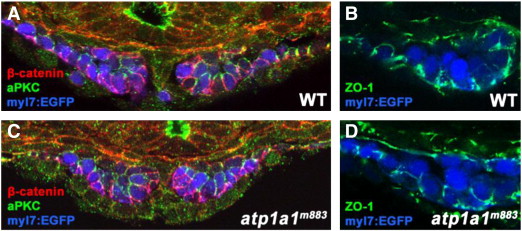
Cell polarity and tight junctions are not disrupted by loss of Atp1a1 function. (A–D) Transverse vibratome sections of 20S stage wild type and atp1a1m883 Tg(myl7:EGFP) embryos. (A,C) The polarized localizations of β-catenin (red, basolateral) and aPKC (green, apicolateral) are indistinguishable between wild type (A) and atp1a1m883 mutant (C) embryos at the 20S stage. (B,D) Tight junctions are present basolaterally in both wild type (B) and atp1a1m883 mutant (D) embryos based on the localization of the tight junction protein ZO-1 (green) at the 20S stage.
Figure Data
Acknowledgments
This image is the copyrighted work of the attributed author or publisher, and
ZFIN has permission only to display this image to its users.
Additional permissions should be obtained from the applicable author or publisher of the image.
Reprinted from Developmental Biology, 362(2), Langenbacher, A.D., Huang, J., Chen, Y., and Chen, J.N., Sodium pump activity in the yolk syncytial layer regulates zebrafish heart tube morphogenesis, 263-270, Copyright (2012) with permission from Elsevier. Full text @ Dev. Biol.