
|
Fig. S6
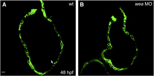
The ventricular myocardium is typically one cell thick at 48 hpf. Single confocal sections display the circumference of the ventricular outer curvature of wild-type (A) and wea morphant (B) embryos at 48 hpf. Tg(myl7:ras-gfp) (Chi et al., 2008) labels cardiomyocyte membranes. In wild-type embryos (A), the ventricular myocardium is typically one cell thick at this stage, and there is little variation in wall thickness ([Chi et al., 2008] and [Peshkovsky et al., 2011]), suggesting that cardiomyocyte volume is generally likely to be proportional to cell surface area at this stage. The same is true in wea morphant embryos (B). Occasional cells bulge toward the lumen of the chamber (arrow, A); this cell behavior seems to occur with a similar frequency in both wild-type and wea morphant embryos. Scale bar is 10 μm.
Reprinted from Developmental Biology, 362(2), Lin, Y.F., Swinburne, I., and Yelon, D., Multiple influences of blood flow on cardiomyocyte hypertrophy in the embryonic zebrafish heart, 242-253, Copyright (2012) with permission from Elsevier. Full text @ Dev. Biol.