
|
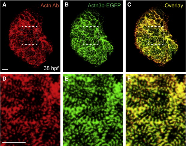
Fig. S2
Tg(myl7:actn3b-egfp) faithfully labels myofibril Z-bands and Z-bodies. (A–C) Ventral view, arterial pole at the top, of a ventricle at 38 hpf from an embryo expressing Tg(myl7:actn3b-egfp). (A) Immunofluorescence (red) localizes α-actinin in Z-bands and Z-bodies within ventricular cardiomyocytes. (B) Native green fluorescence indicates incorporation of the Actn3b-egfp fusion protein into Z-bands and Z-bodies. (C) Overlay and comparison of red and green fluorescence indicates strong correspondence between the incorporation of Actn3b-egfp and the localization of α-actinin. Therefore, the Tg(myl7:actn3b-egfp) transgene provides an effective method for labeling myofibril Z-bands and Z-bodies. (D–F) Higher magnification views of the regions marked by white dashed rectangles in panels A–C. Scale bars are 10 μm.
Reprinted from Developmental Biology, 362(2), Lin, Y.F., Swinburne, I., and Yelon, D., Multiple influences of blood flow on cardiomyocyte hypertrophy in the embryonic zebrafish heart, 242-253, Copyright (2012) with permission from Elsevier. Full text @ Dev. Biol.