
|
Fig. 2
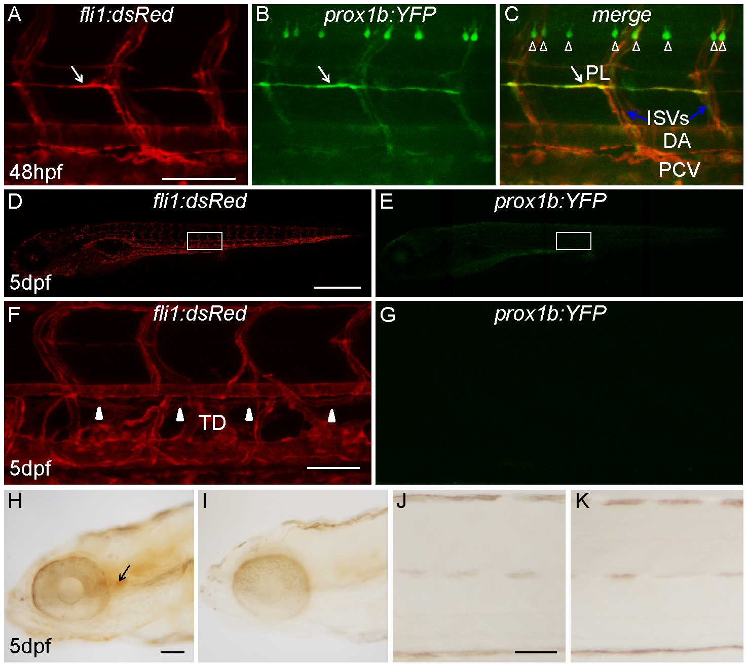
Prox1b does not specifically mark lymphatic aspects of the vasculature.
(A–C) shows prox1b:YFP expression in motor neurons and all endothelial cells of a prox1b BAC:YFP, fli1:DsRed embryo at 48 hpf. White arrows point to parachordal lymphangioblasts. The white open arrowheads label motor neurons. Note that while there is expression of prox1:YFP in PLs, prox1b is also expressed in other (non-lymphatic) aspects of the vasculature. (D) and (E) show the fluorescence images of the same 5-day prox1b BAC:YFP, fli1:DsRed embryo. There is no detectable prox1b:YFP expression in the trunk region of the transgenic embryos (E). (F) and (G) show the enlarged views of the boxed area in (D) and (E). (F) White arrowheads indicate the TD, which resides between DA and PCV. (G) prox1b:YFP expression cannot be detected in TD. (H) and (J) show weak DAB immunostaining against YFP expression in the head (H, indicated by the black arrow), but not in the trunk of transgenic embryos (J). (I) and (K) are DAB staining controls without primary antibody. Scale bars represent 250 μm in (D), and 50 μm in other figures.