
|
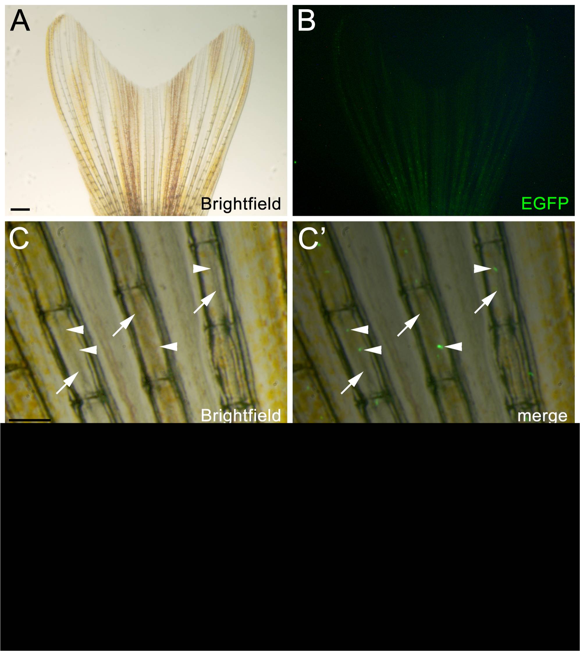
Fig. 3 Wholemount brightfield and flourescent images showing nrd:egfp transgene expression in the adult caudal tail fin.
(A) Brightfield image of the adult caudal fin. (B) Corresponding fluorescent image to panel A. EGFP expression is visualized in the nerve coursing through each bony hemiray of the caudal fin, however at this level of magnification, it is difficult to visualize. (C–C′) High magnification brightfield and corresponding fluorescent overlay showing multiple bony lepidotrichia. The arrows point to the nerve running within each bony hemiray and arrowheads point to EGFP-positive ganglia associated with the nerve. (D and D′) A section of a single bony ray immunolabeled with EGFP to show the transgene and co-labeled with TO-PRO-3 to show all nuclei (magenta). The white line in Panel D shows the location of the nerve. Ep = Epithelium, Bn = bony ray, CT = connective tissue.