
Image
|
Figure Caption
Fig. S1
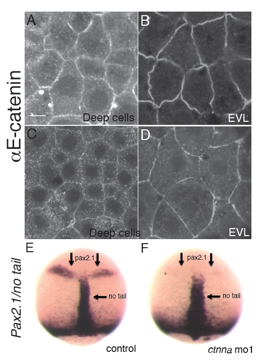
αE-catenin is expressed in deep cells and EVL in early embryogenesis. (A,B) Confocal images of deep cells and EVL cells of embryo at mid-blastula stage stained with an αE-catenin specific antibody. (C,D) Confocal images of deep cells and EVL cells in embryo at 80% epiboly stained with an αE-catenin specific antibody. Scale bar: <10 μm. (E,F) ISH for Pax2.1 and no tail for embryos fixed at 85% epiboly stage. Development of ectoderm (Pax2.1) is delayed in ctnna morphant embryos whereas widened no tail expression is consistent with epiboly delay.
Figure Data
Acknowledgments
This image is the copyrighted work of the attributed author or publisher, and
ZFIN has permission only to display this image to its users.
Additional permissions should be obtained from the applicable author or publisher of the image.
Full text @ Development