
Image
|
Figure Caption
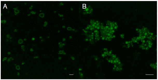
Fig. S1 Fluorescent liposomes are formed following addition of BODIPY-FL analogs to sonicated egg yolk. A. BODIPY-FL C16 partitions preferentially into liposomal membranes making liposomes appear as fluorescent rings when viewed using confocal microscopy. B. BODIPY-FL C5 partitions into the liposomal core, making liposomes appear as fluorescent circles. BODIPY-FL C12, C2, and the BODIPY alone also partition into the aqueous core of liposomes (data not shown). Scale bars are 5 μm.
Acknowledgments
This image is the copyrighted work of the attributed author or publisher, and
ZFIN has permission only to display this image to its users.
Additional permissions should be obtained from the applicable author or publisher of the image.
Reprinted from Developmental Biology, 360(2), Carten, J.D., Bradford, M.K., and Farber, S., Visualizing digestive organ morphology and function using differential fatty acid metabolism in live zebrafish, 276-85, Copyright (2011) with permission from Elsevier. Full text @ Dev. Biol.