
|
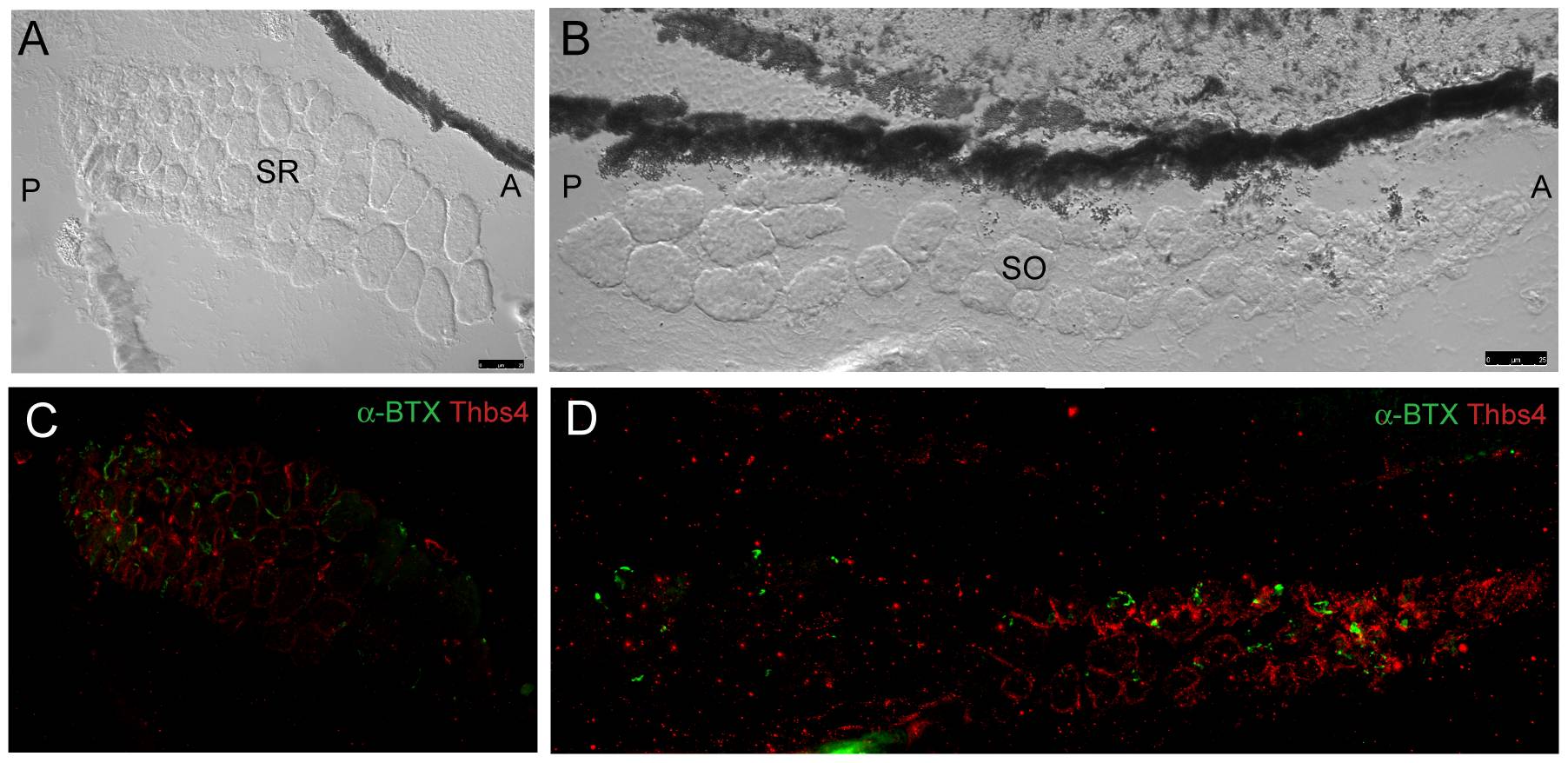
Fig. 10 Fluorescent immunohistochemistry indicates that thrombospondin-4 (thbs-4) localizes to basement membranes of small diameter EOM myocytes.
SR and SO are shown in transverse cross section (A,B – DIC). Thbs-4 (red) localizes to the basement membrane of smaller diameter myocytes and progressively decreases until becoming absent from large diameter myocyte basement membranes (C,D). Smaller diameter fibers appear to be more highly innervated as the density of α-BTX labeled NMJs (green) is greatest in the small diameter myocyte, thbs-4 expressing zone. Images were originally captured with a 40x objective lens and merged together to form mosaics.