
|
Fig. 2
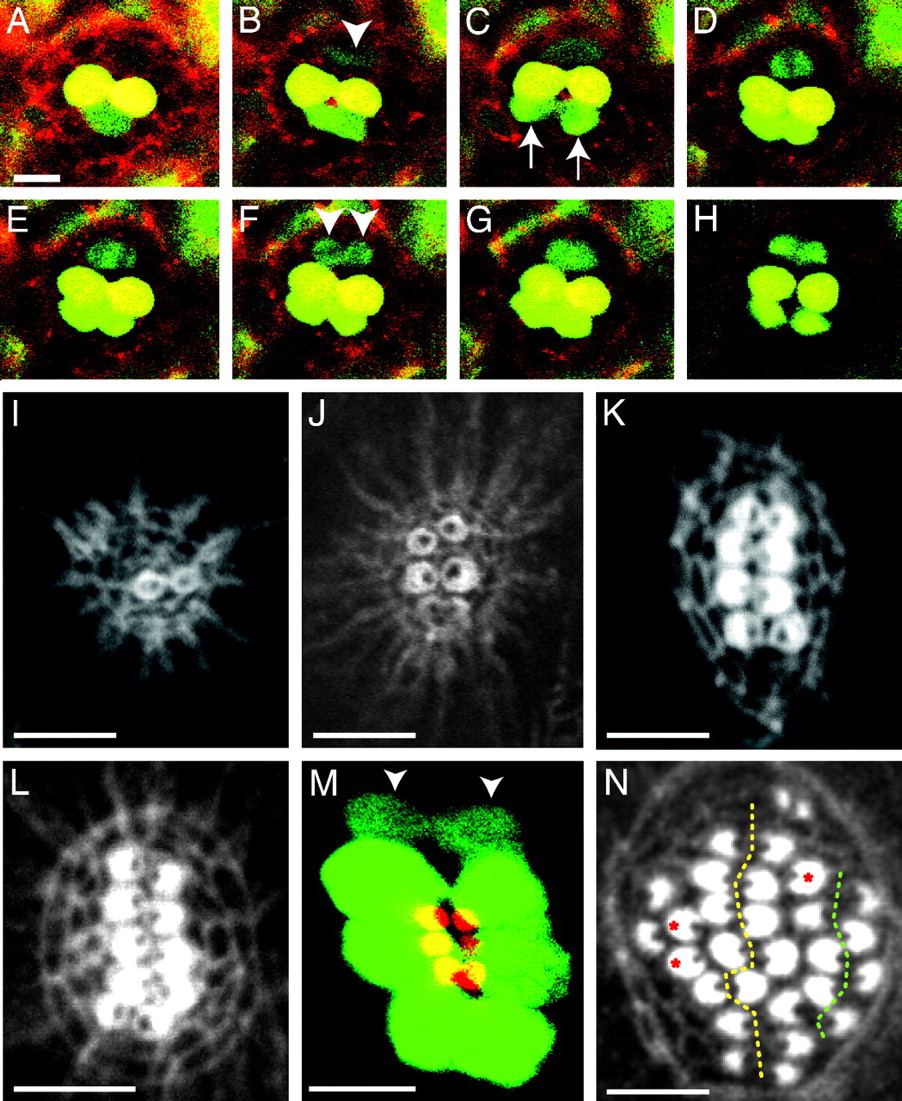
Time-course analysis of hair-cell production. (A–H) In a 12-h-long series of confocal images begun 2 days after fertilization, labeling of an ET4 larva with Texas red-ceramide (red) reveals two GFP-positive mature hair cells (yellow). At the lower edge of the neuromast, a pair of immature hair cells (green; arrows in C), whose proximity initially makes them appear confluent, separate over the course of 3 h (B and C) and become yellow as they mature (D–H). A hair-cell precursor meanwhile develops at the upper edge of the neuromast (arrowhead in B). Over the next hour, this precursor increases its green fluorescence and commences mitosis; the absence of GFP identifies the chromosomes congregated in the metaphase plate (D) and segregated in anaphase (E). The daughter cells (arrowheads in F) eventually separate to form two hair cells (H). This time-lapse series and the supporting movies indicate that hair cells develop in pairs along a single axis in neuromasts. (I) Only 6 h after ablation of hair cells in a wild-type larva, actin staining delineates the first, immature pair of hair bundles. (J) By 10 h after treatment, a neuromast has developed three mature pairs of hair bundles whose opposing polarities are apparent. (K) A neuromast at 16 h after treatment shows the orientation of four pairs of hair bundles. (L) In another animal at the same stage of recovery, five pairs of hair bundles demarcate a line of symmetry dividing the neuromast into two halves along the dorsoventral midline. All of the hair bundles on each side of the line of symmetry have the same orientation. (M) A composite of two confocal images of a regenerating ET4 animal, in which the apical surfaces of the hair cells are pseudocolored red and the cell bodies green, shows that the line of symmetry defined by the hair bundles corresponds to the arrangement of cell bodies. Two immature hair cells have not yet developed hair bundles (arrowheads). (N) By 40 h after treatment, the original line of symmetry is still evident (dotted yellow line), but a second line also has appeared on one side of the neuromast (dotted green line). Three unpaired hair cells are marked with red asterisks. (Scale bars: 10 μm.)