
|
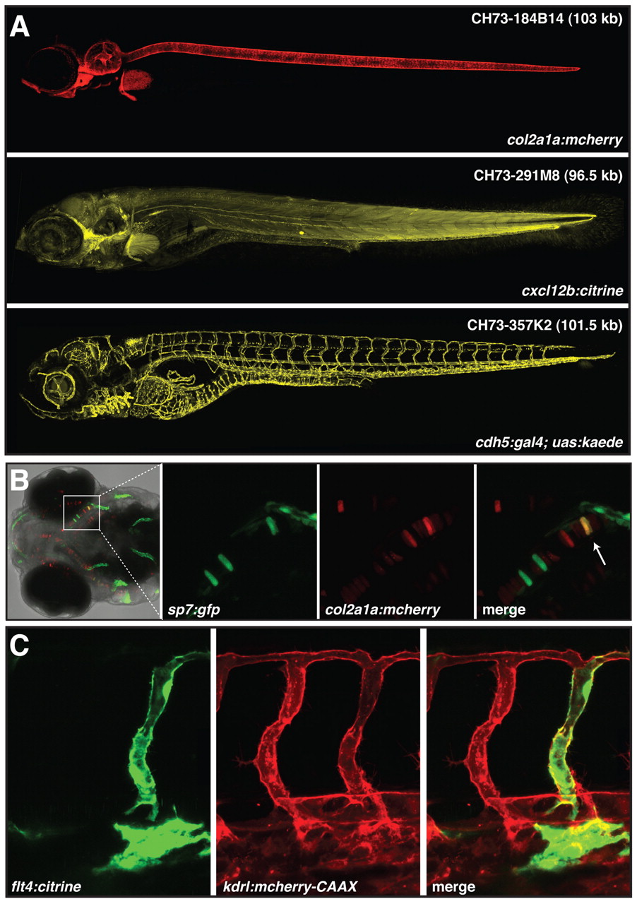
Fig. 3 Colocalization studies using recombineered BAC vectors. (A) Examples of stable, single-copy transgenic lines obtained using the described protocol. Lateral views of whole zebrafish embryos at 4-7 dpf. BAC clone IDs and predicted zebrafish genomic DNA insert sizes are indicated in the top right corner. (B) Dorsal view of the head of an sp7:gfp (Spoorendonk et al., 2008) transgenic zebrafish embryo at 5 dpf injected with col2a1a:mCherry BAC DNA and tol2 transposase mRNA. Note the colocalization of GFP and mCherry in at least one cell (arrow). (C). Lateral view of the trunk of a kdrl:mCherry-CAAX (Hogan et al., 2009) transgenic embryo at 48 hpf injected with a flt4:yfp (citrine) BAC and tol2 transposase mRNA. Individual YFP+ endothelial cells (yellow) are detectable within the posterior caudal vein and intersegmental vessels.