
|
Fig. 5
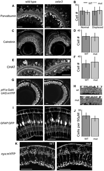
Cell localization and IPL organization are unchanged in the celsr3 mutant.
A) α-parvalbumin labels all displaced amacrine cells and a subpopulation of amacrines in the INL. B) counts of parvalbumin cells, displaced and normal amacrines were counted separately C) α-calretinin labels a subpopulation of amacrines in the INL and all ganglion cells. D) Counts of calretinin positive amacrine cells. E) α-CHAT stains a subpopulation of amacrine cells that laminate in 2 major and 2 minor sublaminae within the IPL. All sublaminae are present in the mutant. F) counts of CHAT positive amacrine cells in the INL G) Tg(ptf1a:Gal4VP16, UAS:mYFP) animals express mYFP in all amacrine and horizontal cells. H) Close ups of the IPL in Tg(ptf1a:Gal4VP16, UAS:mYFP) animals I) Images of Tg(GFAP:GFP) animals, which express GFP in all Müller cells. J) Counts of Müller cells per 50 μM. K) Tg(nyx:mYFP) animals express mYFP in the ON-bipolar cells. Scale bars are 20 μm.