
|
Fig. 5
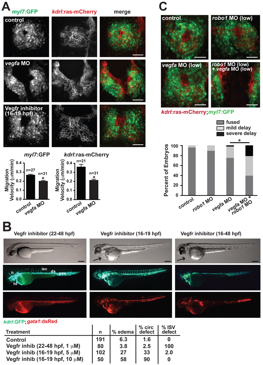
Vegf regulates the migration of the endocardium/myocardium towards the midline. (A) Defects in heart field migration in zebrafish vegfa morphants and embryos treated with Vegfr inhibitor (10 μM Vatalanib at 14-20 somites,16-19 hpf). Quantification of migration velocity of the myocardium [Tg(myl7:GFP) expression] and endocardium [Tg(kdrl:ras-mCherry) expression] in vegfa morphants is indicated beneath. *, P<0.05 compared with control. Data are mean + s.e.m. (B) The patterning of the intersomitic vessels (ISVs) and the head vasculature is severely disrupted by inhibiting Vegfr after heart field migration has occurred (1 μM Vatalanib at 22-48 hpf, left panels), but pericardial edema does not occur and blood flow through the dorsal aorta and posterior cardinal vein is robust. By contrast, edema (arrow) and circulation defects occur when Vegfr is inhibited during heart field migration (10 μM Vatalanib at 16-19 hpf, middle panels), despite the normal patterning of the vessels after removal of the drug at 19 hpf. Continued inhibition of Vegfr (10 μM Vatalanib from 16-48 hpf, right panels) results in profound edema (arrow), which appears to result from severe vascular defects, including a fused dorsal aorta/posterior cardinal vein. Quantification of circulation phenotypes is shown beneath. A circulation defect was defined as reduced blood flow in the axial vessels. (C) Genetic interaction between vegfa and robo1 was assessed by use of sub-phenotypic levels of vegfa and robo1 MOs. The combination of these MOs resulted in profound migration defects observable at the 20-somite stage. Quantification of phenotypes is shown beneath. Mild delay was defined as the contralateral heart fields contacting at the posterior end but remaining unfused anteriorly. Severe delay was defined as a complete absence of heart field fusion at the midline. *, P<0.05 (Fisher′s exact test for a 2×3 contingency table). h, head; ht, heart; isv, intersomitic vessel; da, dorsal aorta; pcv, posterior cardinal vein. Scale bars: 50 μm in A,C; 100 μm in B.