
Image
|
Figure Caption
Fig. 2
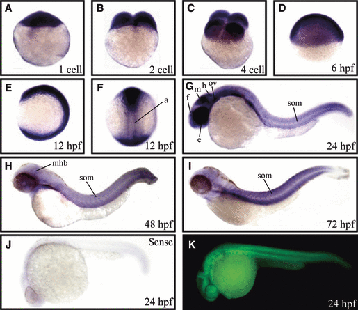
Spatial and temporal expression of prmt1 by WISH and immunofluorescent analysis. Zebrafish embryos at the one-cell stage (A), two-cell stage (B), four-cell stage (C), 6 hpf (D), 12 hpf (E, F), 24 hpf (G), 48 hpf (H) and 72 hpf (I) were analyzed by WISH. A dorsal view of the 12 hpf is shown in (F). WISH with sense riboprobe is shown in (J). Immunofluorescent analysis with anti-PRMT1 of 24-hpf embryos is shown in (K). a, adaxial cells; e, eye; f, forebrain; h, hindbrain; m, midbrain; mhb, mid-hindbrain boundary; ov, otic vesicle; som, somites.
Figure Data
Acknowledgments
This image is the copyrighted work of the attributed author or publisher, and
ZFIN has permission only to display this image to its users.
Additional permissions should be obtained from the applicable author or publisher of the image.
Full text @ FEBS J.