
|
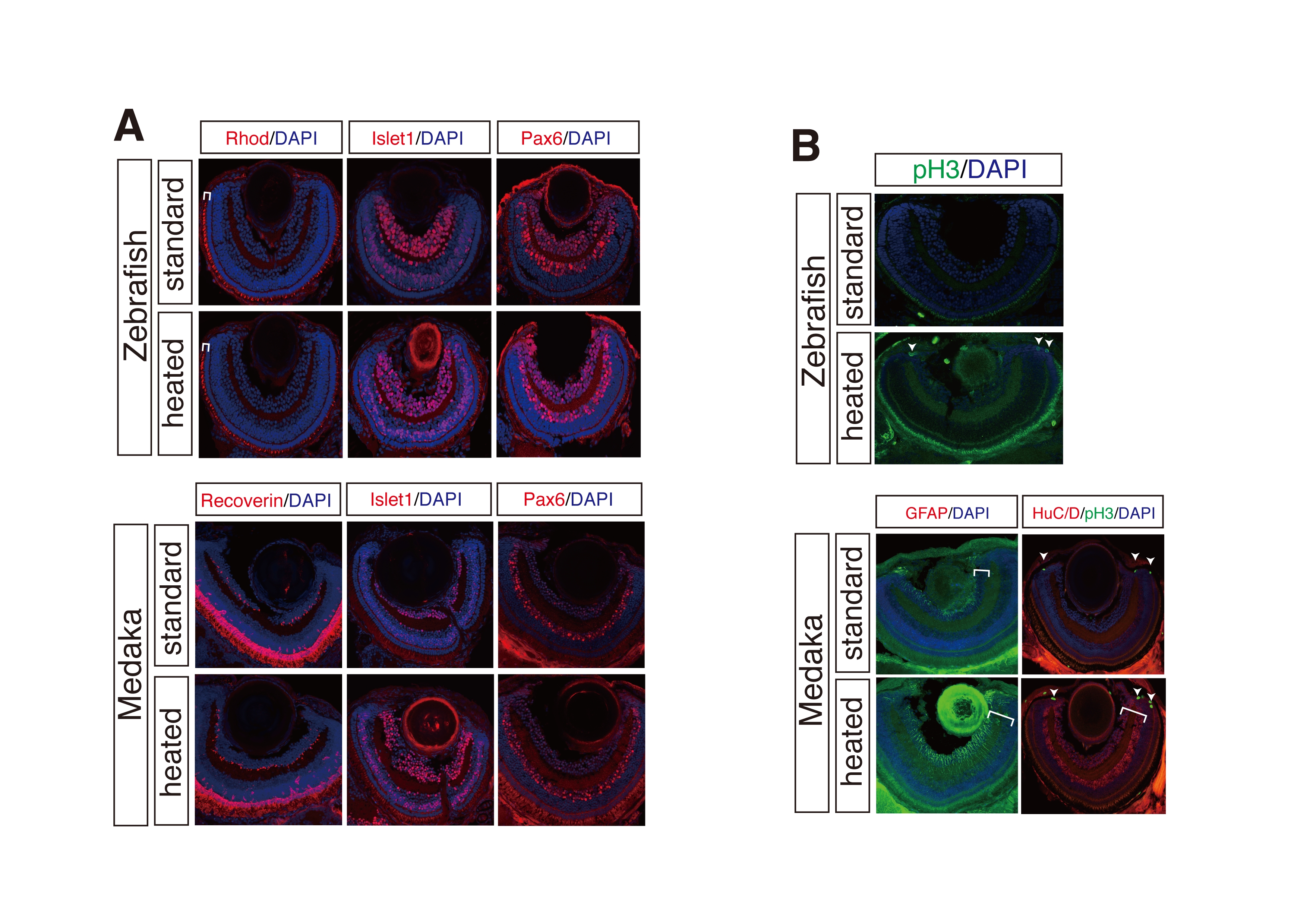
Fig. s1
Comparable and improved fluorescent immunostainings of cryosections by the heating method. (A) Comparable fluorescent immunostainings of cryosections with or without the heating method in zebrafish and medaka. Note that the heating method as well as standard protocol fully preserved the retinal morphology. (B) Immunostainings improved by the heating method. HuC/D (bracket), phospho-histone H3 (pH 3) (arrowheads), and GFAP (brackets) immunostainings were strongly improved as compared to standard protocol in both zebrafish and medaka. (A) and (B) Rhodopsin (brackets) and Recoverin; photoreceptor cell layer. Pax6; amacrine cells. Islet1; retinal ganglion and neuronal cells in the inner nuclear layer. HuC/D; retinal ganglion and amacrine cell layers. pH 3; mitotic retinal progenitor cells. GFAP; Mueller glia cells. Nuclei were counterstained with DAPI (blue).