
|
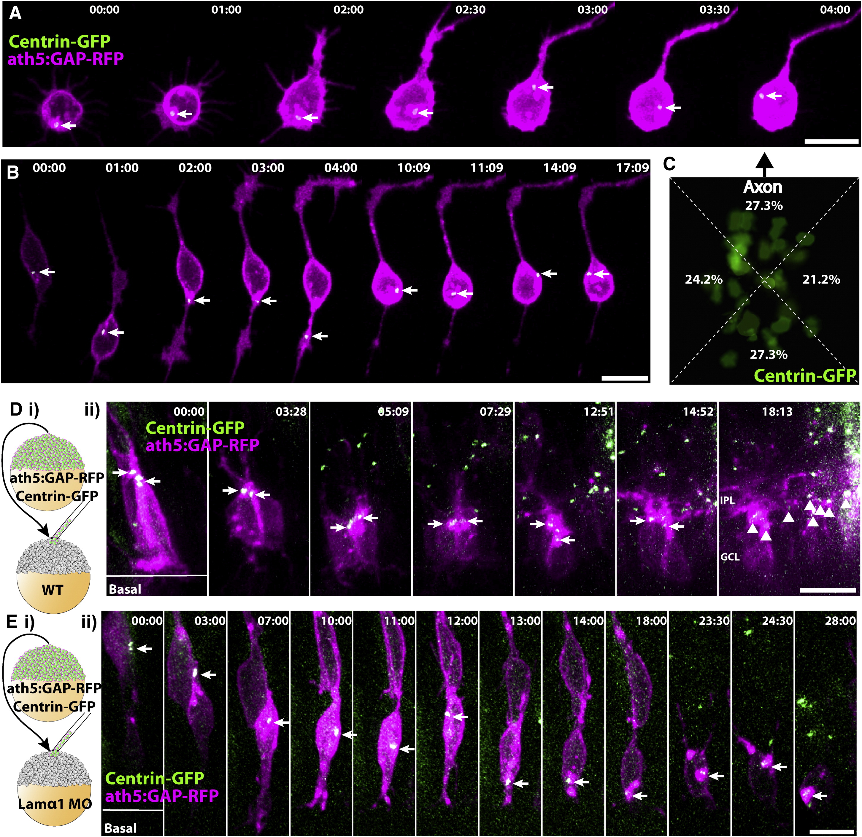
Fig. 4 Centrosomes Are Static and Apically Localized in RGCs Polarizing in WT Retinas, but Mislocalized and Dynamic in RGCs Polarizing In Vitro and in lamα1 Morphants
(A and B) RGCs from ath5:GAP-RFP/Centrin-GFP transgenic embryos were dissociated and imaged during polarization in vitro. Centrosomes (marked by arrow) are not stably localized, but instead move dynamically within the cell body and even within Stage 2 neurites. (C) Overlay of the centrosomes from 33 polarized RGCs in vitro showing their position in reference to the base of the axon (arrow, ath5:GAP-RFP channel not shown). Centroid analysis demonstrates that centrosome location is not biased to any specific quadrant of the RGC cell body (p = 0.9536: Chi square test, n = 33 cells). (Di) Mosaic embryos with WT ath5:GAP-RFP/Centrin-GFP-labeled RGCs in a WT environment were analyzed by time-lapse confocal microscopy. (Dii) Centrosomes (marked by arrows) remain apically positioned in RGCs up until dendrite formation and IPL stratification (marked by arrowheads, t = 18:13). (Ei) Mosaic embryos with WT ath5:GAP-RFP/Centrin-GFP-labeled RGCs in a lamα1 morphant environment were analyzed by time-lapse confocal microscopy. (Eii) The centrosome (marked by arrow) is initially localized apically (t < 07:00), but becomes mislocalized during RGC polarization, and moves dynamically within the cell body. Frames are taken from [Movie S6. Centrosomes Are Dynamic in RGCs Polarizing In Vitro] and [Movie S7. Centrosomes Are Static and Apically Localized in RGCs Polarizing in WT Retinas, but Mislocalized and Dynamic in RGCs Polarizing lamα1 Morphant Retinas]. Time is shown in hr:min; scale bars = 10 μM.