
|
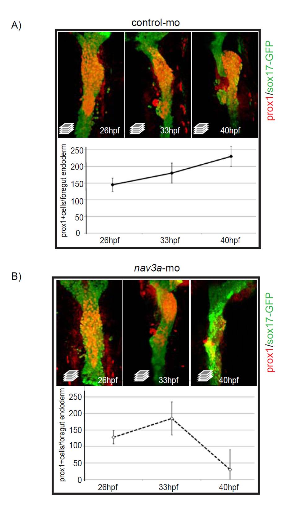
Fig. S10 In nav3a morphant embryos hepatoblasts are correctly specified during early developmental stages. (A,B) Sox17:GFPs870 embryos injected with control morpholino (A) or nav3a-ATG morpholino (B) were analyzed for Prox1 expression in the foregut endoderm by immunofluorescence. During the hepatoblast specification phase between 26 and 33 hpf, the number of sox17-Prox1 double-positive cells is comparable between both groups (images in the left and middle panels, quantification in the graph below the photos). At 40 hpf nav3a morphants failed to form a liver bud. During that outgrowth phase nav3a morphant embryos lost Prox1 expression specifically in the region were hepatoblasts failed to grow out of the foregut endoderm (compare upper right panel in A with upper right panel in B). At 40 hpf the number of Prox1-sox17:gfp double-positive cells is reduced in nav3a morphants. All images are ventral views. Graphs show mean ± s.e.m.