
|
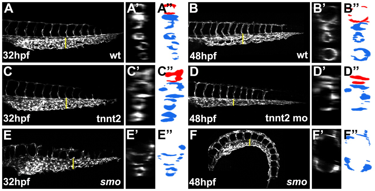
Fig. 4 Blood circulation is not required for sprouting angiogenesis from the caudal vein. (A,B) Caudal vein plexus in wild-type Tg(kdrl:GFP) embryos observed at 32 hpf (A) and 48 hpf (B). (C-F) At 32 hpf, the primordial caudal vein plexus was properly formed in tnnt2 morphant (C) and smo mutant (E) embryos, but the plexus structure was not maintained and degenerated into a single-lumen structure at 48 hpf (D,F). (A′-F′) Optical cross-sections of blood vessels indicated by yellow lines in A-F. (A"-F") Schematic drawings of images shown in A′-F′. Red and blue represent the artery and vein, respectively.