
Image
|
Figure Caption
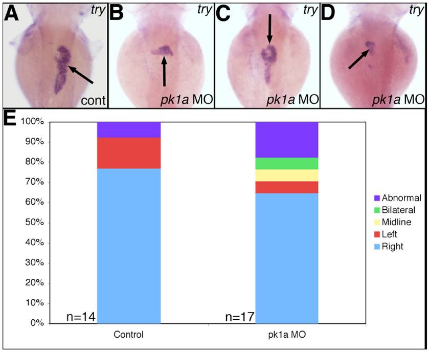
Fig. S4 Marginally abnormal exocrine pancreas localization in pk1a morphants. Whole-mount in situ hybridization of trypsin (try), an exocrine pancreas marker, in 3 dpf control (A) and pk1a morphants demonstrates abnormal exocrine pancreas location, including “bilateral” (B), “midline” (C) and “left” (D). (E) Graph depicting the scoring of pk1a morphants demonstrates a slight difference in localization of the exocrine pancreas (p=NS by chi-square test).
Figure Data
Acknowledgments
This image is the copyrighted work of the attributed author or publisher, and
ZFIN has permission only to display this image to its users.
Additional permissions should be obtained from the applicable author or publisher of the image.
Reprinted from Developmental Biology, 351(2), Cui, S., Capecci, L.M., and Matthews, R.P., Disruption of planar cell polarity activity leads to developmental biliary defects, 229-241, Copyright (2011) with permission from Elsevier. Full text @ Dev. Biol.