
|
Fig. 1
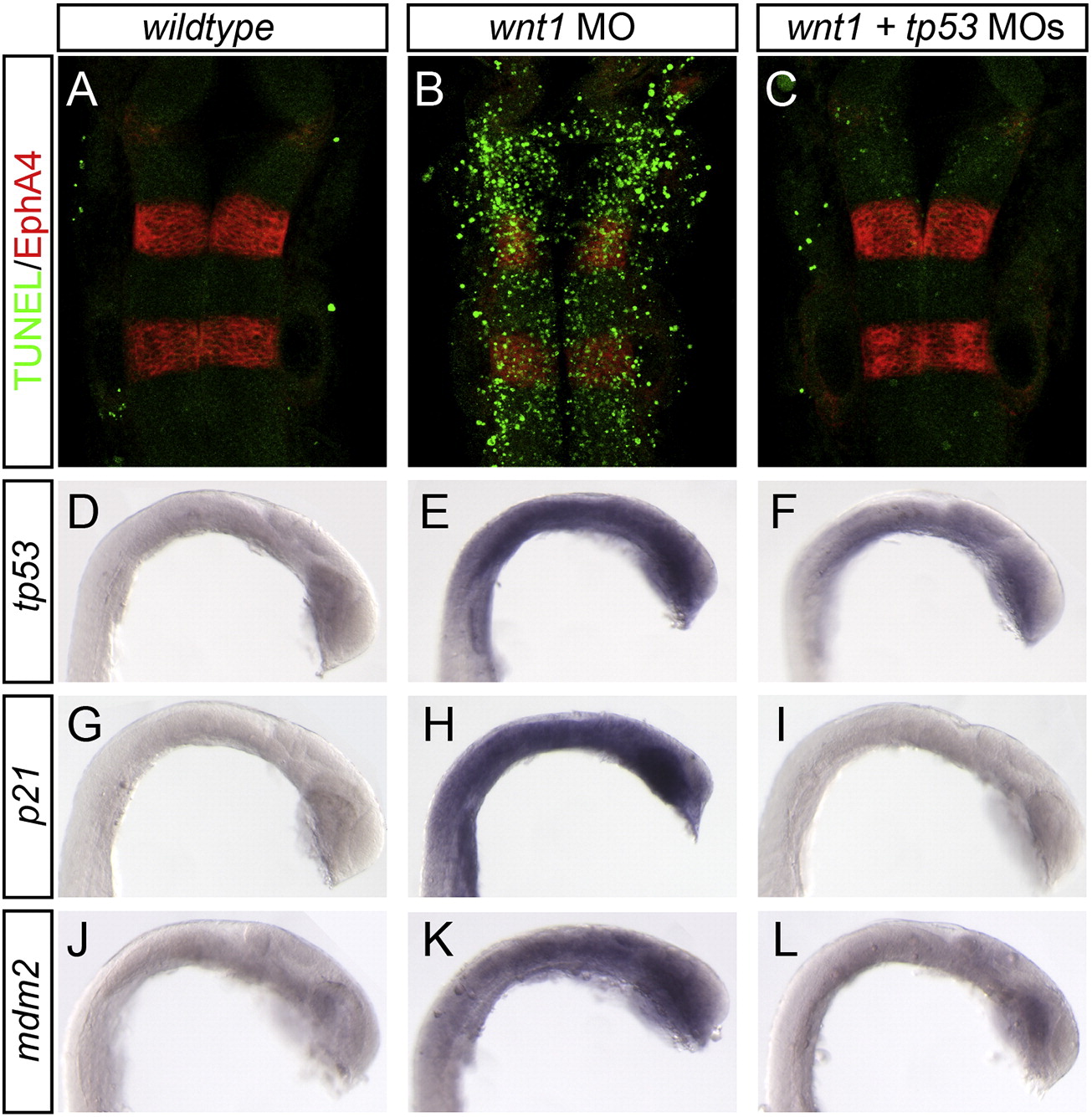
Wnt1 morpholino induces Tp53-dependent apoptosis. Uninjected (A, D, G, J), wnt1 MO-injected (B, E, H, K), and wnt1 + tp53 MO-injected (C, F, I, L) embryos were stained to detect dying cells (A–C, TUNEL, green channel), segment-specific marker EphA4 (A–C, red channel), and Tp53 transcriptional targets tp53 (D–F), p21 (G–I), and mdm2 (J–L). Wnt1 MO-injected embryos show increased cell death (compare A and B, green channel), which is rescued when tp53 MO is co-injected (compare B and C). Tp53 targets are induced in wnt1 MO-injected embryos compared to uninjected controls (compare E, H, K with D, G, J), and this expression is restored to control levels when tp53 MO is co-injected (compare E, H, K and F, I, L).
Reprinted from Developmental Biology, 350(2), Gerety, S.S., and Wilkinson, D.G., Morpholino artifacts provide pitfalls and reveal a novel role for pro-apoptotic genes in hindbrain boundary development, 279-289, Copyright (2011) with permission from Elsevier. Full text @ Dev. Biol.