
|
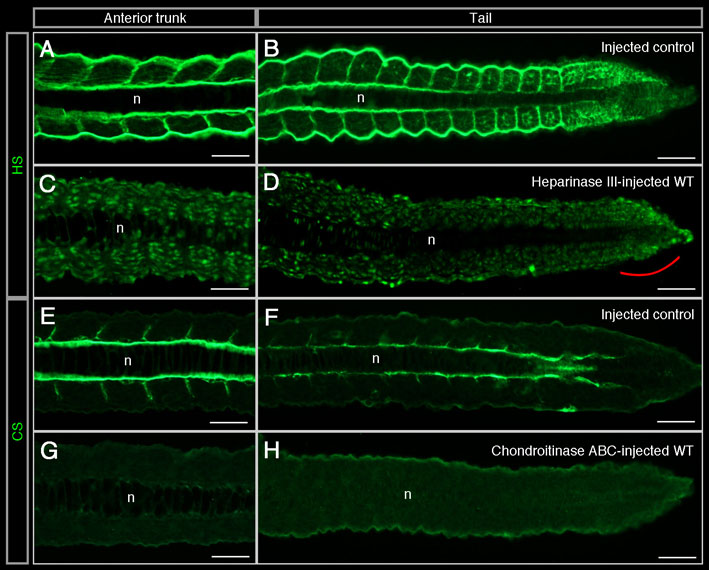
Fig. S2 Heparan sulfate and chondroitin sulfate proteoglycans are removed by heparinase III and Chondroitinase ABC treatments, respectively. (A-H) Frontal sections of vehicle solution-injected wild-type (A,B), heparinase III-injected wild-type (C,D), vehicle solution-injected wild-type (E,F) and Chondroitinase ABC-injected wild-type (G,H) embryos at 24 hpf (i.e. 10 hours post-injection) at the levels of anterior trunk (A,C,E,G) and tail (B,D,F,H). Anterior is on the left. (A-D) In addition to its extracellular labelling, anti-HS antibody (in green) labels all nuclei. Compared with injected controls (n=6/6) (A,B), HSPGs are absent from all heparinase III-injected embryos (n=13/13) (C,D), except for a weak labelling (D, red bracket) in presomitic mesoderm cells (n=13/13) and around the notochord and on the somite surface of the last 7-12 formed somites (n=3/13) (D). The weak labelling in the presomitic mesoderm could be due to de novo synthesis of HSPGs if the enzyme activity declines by 10 hours post-injection or to a HSPGs type that is not degraded by heparinase III. (E-H) Anti-CS antibody is in green. Compared with injected controls (n=7/7) (E,F), CSPGs are absent from the majority of Chondroitinase ABC-injected embryos after a 10-hour treatment (n=7/11) (G,H). A minority (n=4/11) shows a weak labelling at the level of the presomitic mesoderm and last formed somites (H). n, notochord. Scale bar: 50 μm.