
|
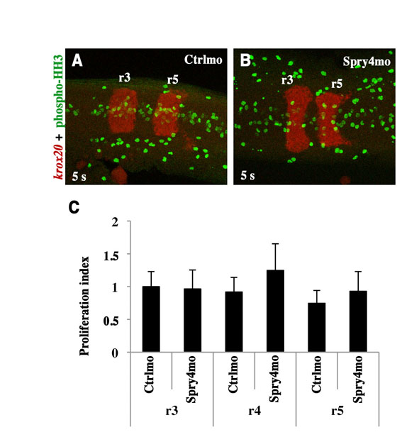
Fig. S4 Cell proliferation in the hindbrain is not affected in Spry4 morphants. (A,B) Immunostaining against phospho-Histone H3 (phospho-HH3, green) was carried out following fluorescent in situ hybridisation using a krox20 probe (red) in 5-somite stage embryos injected with control morpholino (A) or Spry4mo (B). The images are z-projections of nine confocal sections. (C) Normalised proliferation indexes were obtained by dividing the average number of phospho-HH3-positive cells per confocal section in a given rhombomere by the area of the corresponding rhombomere. The ratio obtained in r3 with the control was arbitrarily set at 1, to which the others were then normalised. No significant difference was detected by t-test, P>0.05. Errors bars indicate s.e.m.