
Image
|
Figure Caption
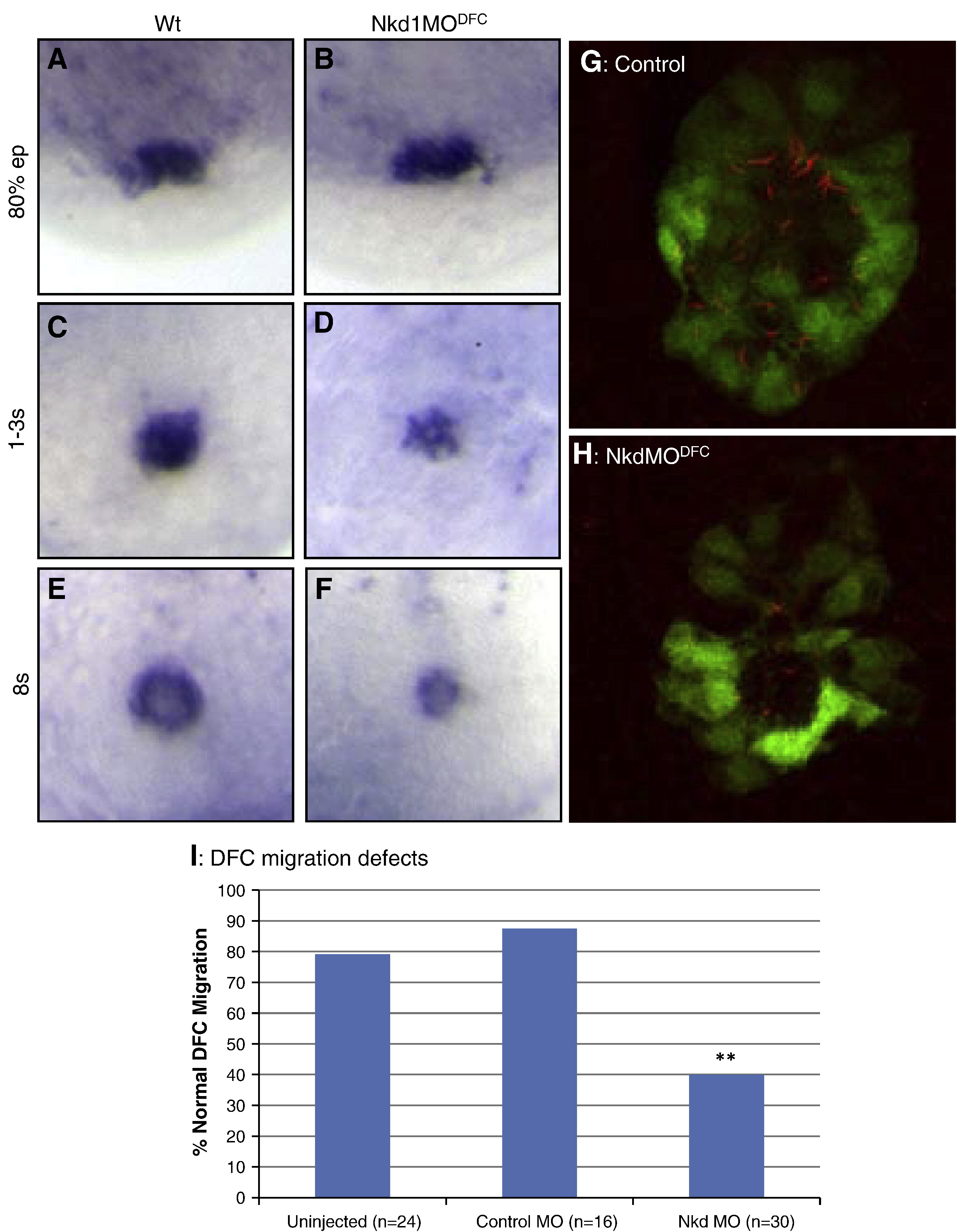
Fig. 3 Nkd1 is required for proper DFC migration and KV formation. (A–F) EGFP (Dusp6:d2EGFP) expression in wt (A, C and E) and Nkd1MODFC (B, D and F) embryos; DFCs were assessed at 80% epiboly (A and B), tailbud to 1-somite stage (C and D) and at 8-somite stage (E and F). (G and H) Fluorescence image denoting KV cilia in wt (J) and Nkd1MODFC (K) embryos. Dusp6:d2EGFP (green) and anti-acetylated tubulin (red) indicate cilia in KV cells. (I) Graph of DFC migration defects. N notes the sample size. ** = p < 0.01 compared to uninjected using Fisher′s exact test.
Figure Data
Acknowledgments
This image is the copyrighted work of the attributed author or publisher, and
ZFIN has permission only to display this image to its users.
Additional permissions should be obtained from the applicable author or publisher of the image.
Reprinted from Developmental Biology, 348(1), Schneider, I., Schneider, P.N., Derry, S.W., Lin, S., Barton, L.J., Westfall, T., and Slusarski, D.C., Zebrafish Nkd1 promotes Dvl degradation and is required for left-right patterning, 22-33, Copyright (2010) with permission from Elsevier. Full text @ Dev. Biol.