
|
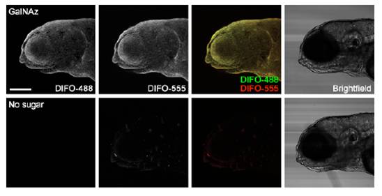
Fig. S9 New azide-containing glycans are presented on the surface of cells out to 96 hpf. Zebrafish embryos were microinjected with 25 pmol of GalNAz (top) or no sugar (bottom) and allowed to develop to 84 hpf, at which point they were reacted with DIFO-488 (100 μM, 1 h). Unreacted azides were immediately quenched by using tris(2-carboxyethyl)phosphine (TCEP) (50 mM, 10 min), and the embryos were allowed to develop further. At 96 hpf, the embryos were reacted with DIFO-555 (100 μM, 1 h) and then imaged by confocal microscopy. Shown are maximum intensity z-projection fluorescence images of surface epithelial cells and corresponding brightfield images. Green, DIFO-488; red, DIFO-555. Scale bar: 200 μm.