
|
Fig. 6
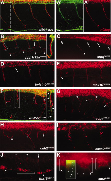
Motor nerve mutants. A-K: Lateral view of the trunk, immunolabeling shows motor axons (green) and motor axon associated glia (red). Anterior left, dorsal up. Confocal z-stacks span the region from the spinal cord exit point to the most superficial location of the spinal nerve. For technical reasons, axon labeling was only successful in ppp1r12a, wnt5bhi1780B, and smo mutants. All other mutants descriptions are based solely on αGfap labeling of motor axon associated glia. A: Wild-type spinal nerves in two full trunk segments. A2,A3: Individual axonal (green) and glial (red) labeling corresponding to the region in A between dashed lines. B: In ppp1r12a mutants motor axons and associated glia terminate (dashed lines) before reaching the lateral line (arrowheads). Motor axons (green) and glial processes (red) are tightly associated (bracket, inset). A mis-localized glial process is seen in one segment (arrow). C-E:sfpq, twistnb, and mak16 mutants had truncated (arrowheads) or absent (arrows) motor axon associated glia. F,G:wnt5b and copa mutants had excessively branched (arrowheads) motor axon associated glia. In some cases motor axons projected correctly (F, brackets, inset, arrowheads), while the associated glia were truncated and disorganized (F,G, arrowheads). H:cdh2 mutants had errors in the positioning of motor axon associated glia akin to motor axon wandering (asterisks), excessive branching (arrowhead) and variable truncations (arrow). I:esco2 mutants had reduced and fragmented motor axon associated glia. J:tbx16 mutants had ectopic glia located ventrally (arrowheads). In some somites glia spanned somite boundaries (bracket). K:smo mutants had ectopic glial clusters (brackets) with aberrant projections (arrows). An overlaid inset shows an over-intensified image of αAT labeling to make visible the rare case in which a motor axon (arrowheads) exits the spinal cord, although is seen here not associated with ectopic αGfap-labeled cell clusters (arrow).