
|
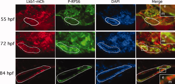
Fig. 6 Co-imaging of Lkb1 localization and TORC1 activation. Zebrafish expressing Lkb1-mCherry (red) at the indicated stages were immunostained for P-RPS6ser240/244 (green). The stages shown represent pre-, mid-, and early post-EIT. At 55 hpf, Lkb1 is diffusely present in the scant cytoplasm of intestinal epithelial cells, though it can be seen in the nuclei of various other cell types outside the intestine. Staining for P-RPS6ser240/244 is low relative to surrounding tissue. At 72 hpf, Lkb1-mCherry is seen in the nuclei of most of the intestinal epithelial cells, and P-RPS6ser240/244 immunostaining is increased relative to 55 hpf. At 84 hpf, nuclear localization of Lkb1-mCherry is sporadically seen in the epithelium, and most (though not all) of those cells stain positive for P-RPS6ser240/244. A white line encloses the gut epithelium. Original magnification, 200×. Insets, 600×. e, epithelium; m, mesenchyme.