
|
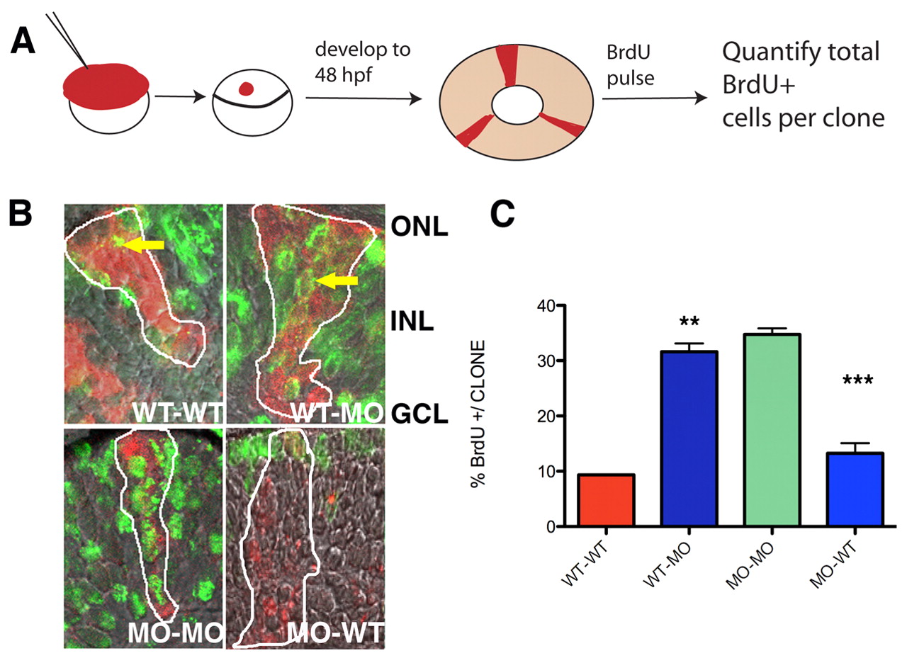
Fig. 9 Id2a downstream function in mediating retinoblast cell cycle progression is non-cell-autonomous. (A) Schematic of experiment. Biotin-dextran lineage-labeled donor cells were transplanted into shield-stage host zebrafish embryos, then mosaic embryos were pulsed with BrdU at 48 hpf to assay cell cycle exit. Clones on the ventral side of the central retina were analyzed. (B) Transplanted cells (red) and BrdU+ cells (green). Top left, wild-type (WT) cells in a WT host; top right, WT cells in an Id2a-MO host; bottom left, Id2a-MO cells in an Id2a-MO host; bottom right, Id2a-MO cells in a WT host. Arrows indicate an example of a BrdU+ cell within a clone. (C) Quantification of the percentage of BrdU+ cells per clone. n=4-5 clones per condition; **, P<0.001; ***, P<0.0003.