
|
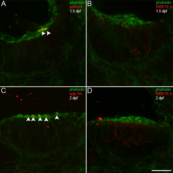
Fig. 7 Complexin 3/4 does not co-localize with RIBEYE b in embryonic inner ear hair cells. (A) A high-magnification confocal projection through the posterior macula in the zebrafish inner ear at 1.5 dpf. Using phalloidin (green) as a marker of developing hair cell stereocilia, one can observe a small number of hair cells with stereocilia. Complexin 3/4 (red) is already apparent among these stereocilia (arrowheads). (B) Dual immunolabeling of RIBEYE b (red) and F-actin with phalloidin (green) in the anterior macula at 1.5 dpf. Note that RIBEYE b has clustered into large puncta on the basolateral membrane of hair cells with stereocilia. (C) At 2 dpf, many more hair cells with stereocilia (green) can be observed in the anterior macula. Complexin 3/4 immunoreactivity (red) can be observed among these stereocilia (arrowheads). (D) RIBEYE b (red) is upregulated in the cytoplasm of more hair cells in the anterior macula at 2 dpf. Scale bar = 10 μm.