
|
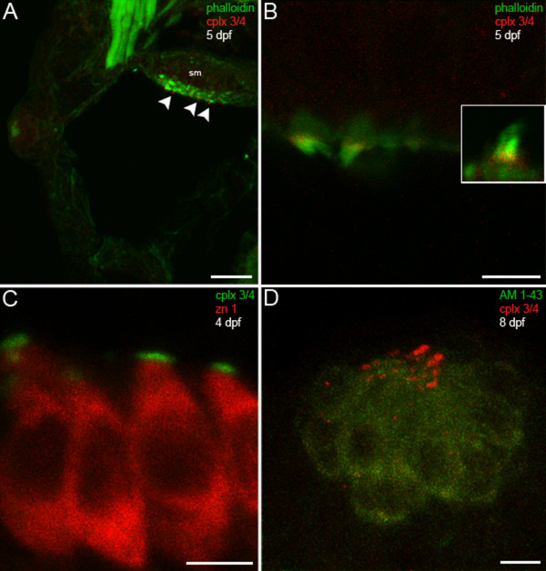
Fig. 6 Hair cells in the larval zebrafish inner ear and lateral line exhibit complexin 3/4 immunoreactivity on their apical surfaces. (A) A confocal projection of a 5-dpf zebrafish transverse section through the otic vesicle reveals complexin 3/4 immunoreactivity (red) on the apical surfaces of inner ear hair cells in the saccular macula (sm; arrowheads) labeled with phalloidin (green). (B) A high-magnification en face view shows complexin 3/4 immunoreactivity (red) at the base of stereocilia labeled by phalloidin above the actin-rich cuticular plate. The inset contains an enlargement of a hair bundle. (C) Complexin 3/4 immunoreactivity (green) directly abuts zn 1 cytoplasmic immunoreactivity (red) on the apical surfaces of inner ear hair cells. (D) To examine neuromast hair cells more closely, larval zebrafish were incubated with AM1-43 (green), fixed, sectioned sagittally, and labeled with anti-complexin 3/4 (red). Complexin 3/4 immunoreactivity is also present on the apical surfaces of neuromast hair cells. Neuromast and inner ear sections incubated with secondary antibodies alone exhibit background immunofluorescence (data not shown). Scale bars: 25 μm (A); 5 μm (B-D).